Find out now about the symptoms and treatment of the most common eye diseases in cats such as conjunctivitis, cataracts and others.
Eye diseases in cats can be caused by a number of reasons: external irritating factors, internal pathologies, hereditary breed predisposition. To minimize the risk of eye diseases, constant care, comfortable conditions for keeping your pet and compliance with preventive measures are necessary.
In this material we have collected information about all common eye diseases. We looked at their causes, symptoms and treatment methods. We have prepared tips for the prevention of eye pathologies.
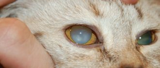
Eyesore (corneal clouding)
A few decades ago, it was much more common to see a person with a noticeable eyesore than it is today. This fact alone should inspire serious and justified optimism, clearly indicating the significant successes of ophthalmology in eliminating this optical and cosmetic defect.
General information
A cataract is any opacity of the cornea; Previously, this concept also included cataract - clouding of the lens - however, taking into account the fundamental etiopathogenetic and anatomical difference, currently these two diseases are separated and considered as independent.
As you know, a healthy cornea is transparent. Being the outermost, open part of the eye's optical tract, the cornea performs both protective and focusing functions. In fact, this is a natural concave-convex lens with a diameter of 1 cm, a thickness of 0.5-1 mm (thinner in the central part, thicker towards the points of diffuse transition into the sclera) and an optical power of 40 diopters.
Any damage, any changes in shape and refractive parameters, degeneration of the multilayer structure of the corneal tissue inevitably and automatically lead to a pronounced and, as a rule, uncorrectable decrease in vision. With all the variety of causes of cloudiness or complete opacity of the cornea, this condition can only be treated surgically, and it is necessary to take all necessary, available, and possible measures to protect the cornea and prevent the formation of a cataract.
Own diseases of the third century
There are a number of specific diseases of the nictitating membrane.
Prolapse (loss) of the lacrimal gland
Lacrimal gland prolapse is rare but occurs in brachycephalic cats. This often occurs during a period of active growth of the cat, at the same time the size of its eyes rapidly increases. The ligament that holds the lacrimal gland of the nictitating membrane in its normal place - under the conjunctiva - is torn. The lacrimal gland emerges into the inner corner of the eye, becomes visible and looks like a small pink rounded formation. When displaced, the lacrimal gland is pinched, it swells and grows in size, and conjunctivitis develops.
Prolapse of the third eyelid lacrimal gland often occurs during rapid growth in cats.
This bothers the cat; when scratching with its paws, secondary flora is introduced, and the course of conjunctivitis becomes purulent. If the lacrimal gland is displaced significantly and for a long time, then its blood circulation begins to suffer and the production of tear fluid decreases. Its pronounced deficiency will lead, in the absence of measures taken, to the development of keratoconjunctivitis sicca. Also against this background, a crease (curvature) of the cartilage of the nictitating membrane may occur.
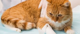
Only surgical treatment is applicable - the displaced lacrimal gland is immersed in the formed conjunctival pocket and sutured with sutures using atraumatic needles and thin absorbable threads (the sutures do not need to be removed later). The operation takes no more than half an hour; in the postoperative period, local and systemic antibacterial drugs are used, as well as an “Elizabethan” collar if the cat rubs its eyes with its paw.
Video: lacrimal gland prolapse
Kink (eversion) of the third eyelid cartilage
The manifestations of a hall of the third eyelid cartilage are similar to the prolapse of the nictitating membrane. Curvature of the cartilage occurs, and part of it is noticeable when examining the inner corner of the eye. In this case, the lacrimal gland can either shift or maintain its normal location. Treatment is also surgical - the curved and protruding part of the cartilage tissue is removed.
The third eyelid cartilage gap can only be corrected surgically
Third eyelid injury
Injury to the third eyelid usually occurs in fights. Initially, there is slight bleeding, secondary conjunctivitis develops, and there may be blepharospasm. Minor injuries heal on their own and without consequences for the function of the nictitating membrane, but in cases where its torn part becomes mobile or cartilage tissue is visualized, a surgical operation is performed to restore the size and full function of the nictitating membrane, as well as eliminate irritation of the conjunctiva by torn tissues and cartilage.
A rupture of the nictitating membrane usually occurs in fights between cats.
Neoplasms of the third century
Neoplasms of the third eyelid are also rare, but are dangerous due to the malignancy of most tumors in this location. A small formation is surgically removed and a histological examination is performed to clarify the nature of the tumor. With a more pronounced spread of the tumor process, the entire nictitating membrane must be removed. The identified type of tumor influences further treatment measures and the prognosis for the cat’s life. Therefore, in all cases of impaired mobility, changes in the structure, shape and color of the third eyelid, it is necessary to exclude the presence of a tumor.
Some veterinarians distinguish lymphoid hyperplasia of the third eyelid - the lymphoid tissue contained in the thickness of the third eyelid grows under the influence of an infectious process or constant irritation; When blinking, the overgrown follicles injure the cornea. The cat has discharge from the eye and blepharospasm. When examined on the surface of the third eyelid, overgrown follicles are identified as a rash or small volumetric formations. Often this causes a similar proliferation of lymphoid tissue on the inner surface of the upper and lower eyelids. Surgical treatment is curettage (scraping) of the overgrown lymphoid tissue, followed by the use of antibiotics and anti-inflammatory drugs.
Causes of corneal opacities
Unlike the lens, which can become cloudy due to endocrine and hematological disorders, to some extent due to the aging of the body and other purely internal, endogenous reasons, the cornea almost never becomes cloudy “on its own.” In the vast majority of cases, the process, injury or other damaging factor that resulted in the occurrence and/or progressive development of the cataract is diagnosed and identified. The main reasons include:
- viral, bacterial, fungal infections, which often begin as “ordinary” conjunctivitis, spread to the cornea (keratoconjunctivitis, keratitis) and, with extensive ulceration, leave an opaque scar surface. As a rule, this is also accompanied by pronounced thinning of the corneal layer and the formation of a convex cataract (ectatic leukoma);
- mechanical wounds and eye damage, incl. trauma during ophthalmic surgery;
- chemical burn of the eye, especially with substances with an intensely alkaline pH reaction;
- intrauterine infectious-inflammatory process resulting in a “ready-made” congenital cataract in the fetus (statistically rare option).
When is a visit to a veterinary ophthalmologist necessary?
Cat owners should be attentive to their pets and if the following signs appear, show them to an ophthalmologist:
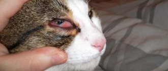
- Swelling and redness of the eyelids.
- The animal constantly scratches its eyes.
- The cat developed photophobia.
- Redness of the eyes.
- Increased tear production.
- Cloudy, purulent discharge from the eyes.
Important!
Do not delay your visit; untimely treatment can lead to partial or complete loss of vision.
Symptoms
Actually, the main and most severe symptom is a decrease in visual acuity in the affected eye. In all cases, the decrease reaches a deep degree, sometimes vision is limited only to residual light sensitivity or complete blindness occurs. With perforations and adhesions (“soldering”) of the cornea and iris, in addition to leukoma, a syndrome of increased intraocular pressure (secondary glaucoma) usually develops.
Depending on the nature of the lesion and its stereometric characteristics, for example, on the degree of convexity of the scar surface, secondary irritation of the conjunctiva of the eyelids may also develop, which creates favorable conditions for recurrent infection. Often, especially when the cornea is thinning and the layer of nerve endings is exposed, patients complain of photophobia, increased lacrimation, and pain.
Recovery
Recovery and prognosis will depend on the severity of the condition and the effectiveness of treatment. Always follow your veterinarian's instructions carefully after treatment or after surgery. It is essential that you take antibiotics for the entire recommended treatment period, even if the condition begins to improve. Failure to do so may result in relapse of the aggressive disease or loss of vision.
Corneal ulcers and keratitis typically begin to heal within three to five days after treatment. Some ulcers caused by infection take longer to heal. Your veterinarian will advise you on your cat's signs.
For cataracts and glaucoma, your veterinarian may schedule follow-up visits as needed to monitor the condition. If cloudy under eyes do not improve or seem to get worse despite treatment, contact your veterinarian immediately.
Treatment of corneal thorn
In cases of exposed nerve endings, severe hyperemia, lacrimation, and painful sensitivity to bright light, special contact lenses-protectors are sometimes prescribed. It should, however, be understood that this is not a treatment and serves only as a palliative method to alleviate symptoms.
Moreover, the countless “folk remedies” that quasi-medical websites vying with desperate patients cannot be considered a treatment: these “recipes” include honey (which for many people is a powerful allergen), aloe, and eyebright in various forms, and mother’s breast milk, and the bile of freshly caught pike, and many other substances, sometimes completely unexpected in this context.
As medicinal support, additional or symptomatic treatment, the ophthalmologist may prescribe an anti-inflammatory regimen, absorbable drugs; the use of physiotherapeutic procedures (for example, electrophoresis with hormone-containing agents) and the prescription of keratoprotectors (Balarpan, Korneregel, etc.) are reported.
However, conservative therapy in this case is just a palliative and does not radically solve the problem. The only effective method of eliminating a cataract has been and remains surgical treatment.
Our ophthalmology center successfully performs surgical treatment of all types of corneal opacities. Using various types of keratoplasty, our ophthalmic surgeons restore vision and remove cosmetic defects. You can find out prices for treatment and make an appointment by calling the phone number listed on the website.
Prevention
If the inner corner of the eyelid has fallen out, it is necessary to urgently show the animal to a veterinarian. Specialists at the White Fang clinic draw attention to the fact that prolapse and protrusion are not separate pathologies, but arise against the background of the progression of other, no less dangerous diseases. The doctor will try to determine why the eye became covered with a third eyelid and prescribe effective treatment.
As a preventative measure, it is recommended to increase the protective functions of the cat’s body, improve nutrition, periodically add vitamins and dietary supplements to the diet, regularly care for the pet’s eyes, systematically undergo a preventive medical examination, and conduct routine deworming. By following these simple rules, you will be able to protect your pet from severe ophthalmic pathologies and preserve vision.
Operations for corneal opacities
Various techniques and protocols are successfully practiced, selected “according to indications” depending on the specific clinical situation. Typically, penetrating or partial keratoplasty is used - respectively, complete (over the entire thickness and area) or partial, more gentle replacement of the affected area or layer of the cornea with a donor keratobioimplant. Such operations are well established and, as a rule, effective, especially if all instructions for the rehabilitation period are followed, which in some cases can last up to a year or more.
According to statistics, the most successful, in terms of short-term and long-term prognosis, are operations for keratoplasty of cataracts caused by infectious ulcerations/scarring. A necessary condition is sanitation and prevention of recurrence of infection.
As for the prevention of the cataract itself, the first and main rule should be to contact a qualified ophthalmologist for any signs of conjunctivitis or other inflammatory process (before the cornea is involved in it), for any injuries and burns of the eye. In a very significant proportion of cases, the cataract could have been prevented without delaying the situation and without bringing the situation to ophthalmic surgery.
Diagnostics: techniques and methods
During the initial examination, the veterinarian evaluates the clinical picture and conducts an external examination of the pet, especially the eye area.
The specialist also collects information about when the spot was first noticed, the quality of food, and studies the animal’s history.
Based on the data obtained, a diagnostic examination is prescribed, including the following manipulations:
- clinical and biochemical blood test;
- ophthalmoscopy;
- serological tests;
- determining the pressure in the eye (to assess how likely it is to develop glaucoma or cataracts);
- Seidel test (fluorescein test);
cytological examination of damaged tissue taken from the conjunctiva;- biopsy;
- microscopic examination of scraping;
- gonioscopy.
If for some reason it is not possible to conduct a standard diagnostic examination, the doctor may prescribe an ultrasound examination, which will help identify the true causes of clouding of the eyes.
If concomitant diseases of viral and infectious origin are suspected, additional diagnostic methods may be prescribed.