Pneumonia is an inflammatory process in the lungs.
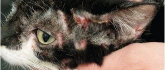
It causes difficulty breathing and lack of oxygen in the blood. This is one of the most common diseases among cats. Although all breeds are susceptible to this disease, brachycephalic (short-faced) cats are more likely to develop upper respiratory tract infections, which can put them at risk for respiratory complications. These breeds include Persians, Ragdolls, Exotics, and Himalayans. Pneumonia is rare in healthy adult cats. Young, old, and sick animals are at increased risk because they have less developed immune systems to fight the virus.
Causes of the disease
The causes of pneumonia in cats are similar to bronchitis. In some cases, the disease occurs as a result of complications after acute bronchitis. The causes of bronchopneumonia include:
• pet hypothermia; • drafts; • getting wet in the rain or sleet; • sleeping on the windowsill; • presence of infectious and invasive diseases; • exposure to harmful gases, dust, smoke and vapors; • ingestion of drugs with the development of aspiration bronchopneumonia; • feeding frozen foods.
Symptoms of pneumonia in cats
When pneumonia occurs in cats, the following symptoms of the disease appear:
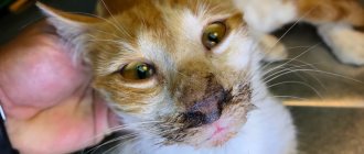
• complete or partial refusal of food; • increased body temperature; • decreased muscle tone; • thirst appears at times; • shortness of breath; • cyanosis of mucous membranes; • depressed state of the cat; • swelling of the paws and chest; • dull, short, painful cough; • purulent nasal discharge with mucus; • slight increase in the border of the lung; • moist rales.
If you have one or more symptoms, you should immediately contact your veterinarian. Remember - it is easier to prevent a disease than to deal with its consequences.
Mycoses of the lungs
Pulmonary mycoses are relatively rare and occur mainly in patients with sharply reduced body defense reactions (AIDS, cancer cachexia, long-term treatment with broad-spectrum antibiotics, glucocorticoids and immunosuppressive therapy). The causative agents of pulmonary mycoses are microscopic fungi (micromycetes). The manifestations of the disease and the nature of its course are significantly different from bacterial and viral infections. Identification of the causative agents of fungal pneumonia is the basis for successful treatment.
Aspergillosis
Invasive aspergillosis (pneumonia). The main causative agents of invasive aspergillosis are: A. fumigatus (60-90%), A. flavus (10-30%) and A. niger (2-15%). The frequency is 12-34 cases per 1 million population per year. Infection occurs through inhalation of Aspergillus conidia. Aspergillosis is not transmitted from person to person. It most often develops in patients with acute leukemia during cytostatic therapy, in patients receiving glucocorticoids and immunosuppressants for a long time. Clinical manifestations The duration of the incubation period is not determined. The most common signs of the disease are ineffectiveness of broad-spectrum antibiotics, fever over 38°C lasting more than 96 hours, nonproductive cough, chest pain, hemoptysis and shortness of breath. In 30% of cases, no increase in temperature may be observed. Sometimes the clinical picture of aspergillosis resembles signs of thrombosis of the branches of the pulmonary artery: sudden chest pain and severe shortness of breath. Often the only manifestation of the disease is changes on an x-ray or CT scan of the lungs.
Diagnostics
Main detection method:
- lesions - multislice computed tomography (MSCT);
- microbiological confirmation of the diagnosis - microscopy and culture of respiratory substrates;
- serological diagnosis - determination of Aspergillus antigen (galactomannan) in blood serum.
Diagnostic methods:
- radiography of the lungs and paranasal sinuses;
- Chest CT (MSCT);
- in the presence of neurological symptoms - MSCT or MRI of the brain;
- determination of Aspergillus antigen in blood serum;
- bronchoscopy, bronchoalveolar lavage (BAL), biopsy of lesions;
- microscopy and culture of BAL swabs, sputum, nasal discharge, and biopsy material.
Treatment includes antifungal (antifungal) therapy, elimination of risk factors, and surgical removal of affected tissue. Forecast. Without treatment, invasive aspergillosis almost always ends in death (within 1-4 weeks). During treatment, mortality is 30-50% and depends on the underlying disease (acute leukemia, etc.), as well as on the prevalence of aspergillosis or localization of the disease (dissemination, central nervous system damage)
Chronic necrotizing pulmonary aspergillosis is a relatively rare disease, accounting for approximately 5% of all cases of pulmonary aspergillosis. The causative agent is A. fumigatus, less commonly - A. flavus, A. terreus, etc. Risk factors are AIDS, diabetes mellitus, alcoholism, long-term treatment with steroids. Symptoms are characteristic, but not specific. Usually a chronic productive cough (with sputum) develops, often with moderate hemoptysis. Low-grade fever. General weakness and weight loss. The disease occurs “chronically” with periodic exacerbations and progressive impairment of lung function due to the development of fibrosis. Complications: damage to the pleura, ribs, vertebrae, pulmonary hemorrhage, invasive pulmonary aspergillosis (pneumonia) with hematogenous dissemination, damage to the brain and internal organs. Diagnosis - see invasive aspergillosis. Treatment. Long-term use of antifungal drugs. The indication for surgical treatment is a high risk of pulmonary hemorrhage.
Aspergilloma (non-invasive aspergillosis) , or “mushroom ball”, is the mycelium (mycelium) of the Aspergillus fungus that grows in the cavities of the lungs formed as a result of tuberculosis, tumors and other diseases. Aspergilloma can occur in the paranasal sinuses. The causative agent is Aspergillus fumigatus, less commonly A.flavus. Tuberculosis is the cause of cavity formation in aspergilloma in 40-70% of cases; destructive pneumonia - in 10-20%; bullous emphysema - in 10-20%; bronchiectasis - in 5-10%; tumors - in 3-7% of cases. The probability of developing aspergilloma in a cavity measuring 2 cm is 15-20%. Usually occurs between the ages of 40 and 70, more often in men. Initially, it is asymptomatic, but as it progresses, coughing begins to bother you, hemoptysis, and low-grade fever occur. With secondary bacterial infection of the cavity affected by fungi, signs of acute inflammation may develop. In most cases, aspergilloma occurs in the upper lobe of the right lung (50-75%), less often - in the upper lobe of the left lung (20-30%). In approximately 10% of patients, signs of aspergilloma resolve spontaneously, without treatment. During the course of the disease, most patients experience an episode of hemoptysis, and 20% have pulmonary hemorrhage. Complications of aspergilloma include pulmonary hemorrhage and invasive growth of Aspergillus with the development of chronic necrotizing pulmonary aspergillosis or specific pleurisy. Diagnosis - see invasive aspergillosis. Treatment is carried out in case of development or high risk of complications (repeated hemoptysis, pulmonary hemorrhage, etc.); observation is indicated for asymptomatic aspergilloma. The main treatment method is surgical removal of the affected area of the lung.
Allergic bronchopulmonary aspergillosis is characterized by the development of a type I hypersensitivity reaction when the respiratory tract is affected by Aspergillus. The frequency in patients with bronchial asthma is 1-5%, in patients with cystic fibrosis - 5-14%. Pathogens - Aspergillus fumigatus, A. clavatus, less often - other Aspergillus. Congenital predisposition contributes to its occurrence. No invasive damage to lung tissue is observed. The disease usually occurs chronically with periodic exacerbations of broncho-obstructive syndrome and/or the occurrence of eosinophilic infiltrates. The main signs of exacerbation are attacks of suffocation, increased body temperature, chest pain and cough with sputum containing brown inclusions and mucus plugs. Over a long period of time, bronchiectasis and pulmonary fibrosis develop, leading to respiratory failure.
- Invasive candidiasis
- Pulmonary cryptococcosis
- Zygomycosis of the lungs
- Hyalohyphomycosis
Invasive pulmonary candidiasis (candidal pneumonia) is relatively rare and accounts for approximately 5-15% of all cases of invasive candidiasis. It can be primary or secondary, resulting from hematogenous dissemination of Candida from another lesion. It develops mainly in patients against the background of severe pathology (surgical treatment of the gastrointestinal tract, infected pancreatic necrosis, long-term parenteral nutrition, the use of immunosuppressants, artificial ventilation, hemodialysis, repeated blood transfusions, diabetes mellitus). The main causative agents of candidal pneumonia are C. albicans, C. tropicalis, C. parapsilosis, C. glabrata, etc. Many Candida are inhabitants of the human body and are detected by culture from the mucous membrane of the oral cavity and gastrointestinal tract in 30-50% of healthy people . Clinical signs (fever, nonproductive cough, shortness of breath, and chest pain) are nonspecific and do not distinguish candidal pneumonia from bacterial or other pulmonary mycosis. The basis of diagnosis is the identification of Candida during histological examination and/or culture of a lung biopsy. Treatment. An important condition for successful treatment is identification of the pathogen. Candida species clearly correlates with sensitivity to antimycotics.
The incidence of cryptococcosis has increased significantly in recent years due to the HIV pandemic. In the vast majority of cases, the causative agent of cryptococcosis is Cryptococcus neoformans, which is distributed everywhere (in soil, on plants, in bird feces). The main risk factors are disorders of cellular immunity caused by AIDS, lymphoma, chronic lymphocytic leukemia, T-cell leukemia, as well as long-term use of glucocorticoids and immunosuppressants. The risk of developing cryptococcosis is determined by the severity of immunodeficiency. For example, the incidence of cryptococcosis in AIDS patients is up to 30%. Cryptococcosis rarely develops in immunocompetent patients. It occurs less frequently in women than in men, and less frequently in children than in adults. Infection occurs by inhalation. In patients with AIDS, the central nervous system, lungs, and skin are most often affected; disseminated variants of infection develop, involving the bones, kidneys, adrenal glands, etc. The main signs are fever (81%), cough (63%), shortness of breath (50%), weight loss (47%), rarely - chest pain and hemoptysis. Diagnosis: see invasive aspergillosis. For any localization of cryptococcal infection, a lumbar puncture is necessary (determination of intracerebral pressure, microscopy of cerebrospinal fluid and culture for flora). Treatment. The choice and duration of use of antimycotics are determined by the patient’s condition and the localization of the process.
Pathogens are lower fungi belonging to the class Zygomycetes. Zygomycetes are ubiquitous, live in the soil, and are often found on food products and in rotting plant waste. The pathogen enters the lungs by inhaling spores. The risk factors are the same as for other mycoses. Zygomycosis is characterized by an extremely aggressive course with very rapid destruction of all tissue barriers, damage to blood vessels, hematogenous dissemination with the subsequent development of thrombosis, infarction and tissue necrosis. With zygomycosis, any organs can be affected, but the paranasal sinuses (35-50% of all cases), lungs (20-30%), skin and subcutaneous tissue (10%), as well as the gastrointestinal tract (5-10%) are most often affected. %). Pulmonary zygomycosis is usually manifested by an increase in body temperature of more than 38 ° C, which does not decrease during treatment with broad-spectrum antibiotics, cough, chest pain, profuse hemoptysis or pulmonary hemorrhage.
Hyalohyphomycosis is a group of diseases caused by the fungi Fusarium spp., Acremonium spp., Paecilomyces spp., Scedosporium spp., Scopulariopsis brevicaulis and Trichoderma longibrachiatum. The pathogens are ubiquitous, often found in the soil and on various plants.
Fusarium is considered the second most common causative agent of invasive pulmonary mycoses after Aspergillus. In addition to pneumonia, hyalohifomycetes cause local lesions in immunocompetent patients, and fungemia (fungus in the blood) and disseminated infections, which are characterized by very high mortality, in immunocompromised patients. The unfavorable prognosis is associated both with the severity of immunosuppression in patients and with the low sensitivity of hyaloghiphomycetes to most used antimycotics.
Hyalohyphomycosis of the lungs most often develops in patients with hemoblastosis or recipients of bone marrow transplants, much less often in patients with widespread deep burns. Infection usually occurs by inhalation. One of the possible sources of the pathogen is the affected nails with onychomycosis. Pathogens can infect arteries with subsequent development of thrombosis, infarction and hematogenous dissemination. The disease usually begins as pneumonia or sinusitis, and as it progresses, hematogenous dissemination develops with damage to the skin, internal organs, bones and brain. The clinical picture of the disease is determined by the localization of the process; a common symptom is fever refractory to antibiotics. In 55-70% of patients, characteristic lesions of the skin and subcutaneous tissue develop: painful erythematous papules or subcutaneous nodules followed by the formation of a focus of necrosis in the center.
Diagnosis: see invasive aspergillosis.
Treatment. The causative agents of hyalohyphomycosis are characterized by low sensitivity and even resistance to antimycotics
Diagnosis of pneumonia in cats
In order to diagnose pneumonia and begin treatment for a cat, you need to go not just to a veterinarian, but to a clinic equipped with a laboratory. There is a completely logical explanation for this, because making a diagnosis, identifying the form and treating it requires not only information received from the pet owner, but also a thorough examination. For example, eosinophilic pneumonia is detected when a large number of eosinophils are detected in the blood fluid. In this case, treatment should focus on treating not the pneumonia, but the parasites in the body. In general, when visiting a veterinary clinic with symptoms of pneumonia in a cat, the following must be done:
• study of anamnesis; • examination of the cat; • fluoroscopy; • microscopy of mucus; • collection of information about the epizootological situation of the site or settlement.
Diagnostics
To identify the disease, the doctor examines the pet and asks the owners for complaints. To identify pneumonia, a physical examination is performed and a referral is given to the laboratory and to the instrumental diagnostic room.
The animal is prescribed:
- auscultation - listening to the lungs;
- taking blood tests;
- radiography or ultrasound.
Based on the results of the examination, an accurate diagnosis is made, indicating the type of pneumonia and the extent of the pathological process. Next, the doctor decides whether to hospitalize or prescribe outpatient treatment.
Treatment of pneumonia
Treatment is prescribed by a veterinarian depending on the form of the disease and the symptoms present. As a complex therapy it is recommended:
• balance the cat's diet; • provide the animal with the necessary vitamins and microelements; • applying heat to the chest; • antibiotics, sulfa drugs; • UHF; • inducotherapy; • lacto- and autohemotherapy; • immunoglobulins; • salicylic drugs; • diuretics; • expectorants; • blockades.
Prevention of pneumonia, favorable conditions after hospitalization
If the diagnosis of pneumonia is confirmed, your pet will most likely be hospitalized for several days. Treatment includes antibiotics, penicillin or amoxicillin, and diuretics to clear accumulated fluid in the airways.
After discharge, prepare a bright, ventilated room for the cat. Always heat food and water until warm. A proper diet rich in vitamins and minerals is the key to health! Bring the food to a liquid pulp, so it will be more convenient for the cat to eat. If Fluffy refuses to eat, try his favorite canned food.
Keep your cat warm, isolating him from other pets, so as not to unnecessarily irritate your pet. Do everything to ensure that he gets as much rest and sleep as possible.
Regularly visit a veterinary clinic for preventive examinations, consult with a doctor and revaccinate.
Sign up for treatment for pneumonia
POLAR BEAR
The veterinary clinic in Nizhny Novgorod "White Bear" provides all types of services for cats and dogs.
Conducts any examinations and tests in its own veterinary laboratory; the clinic operates a veterinary ambulance for animals and a 24-hour veterinarian is on call at home. Agreement on the processing of personal data
Public offer
SITE MENU
- About the clinic
- Price list for veterinary clinic services
- Treatment
- Services
- Price list for veterinary clinic services
- Vetapteka
- Contacts
- Site Map
CONTACTS
- Nizhny Novgorod st. Vyatskaya 7
- 8 (831) 437-25-27
- [email protected]
What are the dangers for cats with pneumonia?
Pneumonia is not a localized inflammatory process, but a disease of the entire body as a whole. As a result of the resulting inflammation, various consequences are noted:
• hyperemia and pulmonary edema; • hemorrhages; • proliferation; • atelectasis; • exudation; • poisoning of the body with toxins; • necrosis; • reduction of the respiratory surface of the lungs; • increased respiratory movements; • reduction of oxygen consumption; • reduction of gas exchange in the body; • violation of protein, fat, carbohydrate and vitamin-mineral metabolism; • development of cardiovascular failure.
In the gastrointestinal tract
Klebsiella forms colonies in the intestines after birth in humans during the first week of life. This is normal and no treatment is required. Typically, about 105 bacteria are isolated in 1 g of feces. This indicator increases with disorders or diseases, infection of the gastrointestinal tract. Taking antibiotics leads to the suppression of pathogenic flora and normal flora needed by a person. Saprophytes with pathogenic effects actively reproduce. Clabsiosis in the intestines appears as a result of:
- infections from carriers;
- dysbacteriosis.
The patient progresses gastroduodenitis, gastritis, and intestinal infection with an abrupt onset and severe symptoms. The duration of the severe condition is up to 5 days.
Klebsiella often becomes the cause of nosocomial infections, when mass infection of all patients and staff with antibiotic-resistant strains occurs. Severe damage is observed with weak immunity and chemotherapy courses.
The described pneumonia bacterium causes lesions of the genitourinary tract, because for them it always remains a pathogenic flora. It often affects the vaginal environment, especially if it undergoes a course of antibiotic therapy that disrupts the normal microflora.
Klebsiella rhinoscleroma, also known as the Frisch-Wolkovich bacillus, deserves special attention. It entails the development of scleroma as an infectious disease that affects the upper respiratory tract and the mucous membrane of the nose and bronchi. Infiltrates appear followed by scarring.
As for Klebsiella ozena, it is found in 80% of patients with diagnosed ozena, the so-called foul runny nose. The disease is accompanied by atrophy of the bone walls and mucous membrane in the nose, which causes a secretion to appear that, when dried, produces foul-smelling crusts. This bacterium leads to chronic damage:
- respiratory system;
- trachea;
- larynx;
- throats.
Preventive measures
To prevent pneumonia in cats, it is recommended to protect pets from the following unfavorable factors:
• dampness; • smoke; • dust; • drafts; • aniline paints; • exposure to harmful vapors of acids and alkalis.
In addition, it is worth paying attention to a number of actions that are essential to follow in order to maintain the health of the animal for as long as possible and in the best possible way.
- The animal must be vaccinated in a timely manner against viruses that can cause pneumonia, both primary and secondary. Experts believe that vaccination should be done not only for cats that have access to the street, but also for pets.
- After contact with other animals, upon arrival home, a person must wash their hands and change clothes before petting a pet. Often the virus can be transmitted by airborne droplets.
- The room in which the animal lives must be cleaned regularly, using special disinfectants. This will prevent the formation and spread of bacteria and fungus.
Attention: the above is for educational purposes only and does not constitute professional medical advice or scientific material.