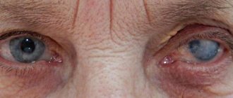
A cat's health can be judged with confidence by its appearance. The organs of vision are the first to react to its deterioration. The eyes of a healthy cat are bright, clean, without traces of discharge in the corners. And if their condition is far from ideal, we can assume that the animal is sick.
Red eye syndrome in cats is a very common symptom. It often indicates ophthalmological problems, and sometimes indicates a somatic disease. Reddened protein in most cases is accompanied by itching, burning, lacrimation, photophobia, hemorrhages in the iris, and blepharospasm. Red, inflamed eyes indicate that blood circulation in their choroid is impaired, the blood vessels are dilated, and the surrounding tissues are inflamed. From this article you will learn what causes eye redness and what to do to help your cat.
Diagnostics
Your veterinarian will perform a complete physical examination of your cat, including a blood chemistry profile, complete blood count, urinalysis, and electrolyte analysis.
You will need to provide details about your cat's health, the occurrence of its symptoms, and possible incidents that may have triggered the condition.
Red eyes are often a visible sign of an underlying systemic disease, sometimes serious. Therefore, a blood test is necessary to exclude or confirm the underlying disease.
To rule out cancer and infectious causes of red eyes, X-rays can be used to visually examine the chest and abdomen.
Equally useful for diagnostic purposes are ultrasound images of the eye, which can be performed if the eye is opaque, and tonometry, which is the measurement of pressure inside the eye using a tonometer.
If there is purulent discharge from the eye or long-standing eye disease, your veterinarian will perform an aerobic bacterial culture and sensitivity profile.
Other tests your veterinarian may choose are the Schirmer tear test, used to check for normal tear production; cytological (microscopic) examination of cells of the eyelid, conjunctiva and cornea; and a conjunctival biopsy (tissue sample) if there is chronic conjunctivitis or mass lesions.
For cats, a polymerase chain reaction (PCR) test may be used to diagnose an inherited or infectious disease, or an indirect fluorescent antibody (IFA) test of corneal or conjunctival scrapings may be used to test for herpes virus and chlamydia bacteria. both of which can indirectly affect the eyes.
Fluorescein corneal staining, which uses a non-invasive dye to coat the eye making abnormalities more visible under light, can also be used to detect foreign material, ulcers, scratches and other damage on the surface of a cat's eye. .
Eye diseases causing redness
The following diseases can contribute to the appearance of such a symptom:
- Blepharitis. The disease is accompanied by inflammation of the eyelid skin caused by bacteria or fungi. May occur due to an allergic reaction or injury. Blepharitis is accompanied by swelling and baldness of the eyelids, squinting, frequent blinking, itching, mucous or purulent discharge.
- Conjunctivitis. The disease is of viral or bacterial origin. In the latter case, the inflammatory process is characterized by an abrupt onset and rapid course. The clinical picture of viral conjunctivitis develops more slowly. In this case, purulent discharge does not always appear. Liquid mucus may be released from the conjunctival sac, which does not stick the eyelids together after sleep. This makes diagnosis very difficult. In the bacterial form of inflammation, thick purulent discharge sticks the eyelids together. The redness is mild.
- Glaucoma. The disease is characterized by an increase in intraocular pressure. It is diagnosed frequently. The sclera in cats turns red due to vasodilation. An enlarged pupil does not respond well to light. The cat tries to stay in dark rooms. The cornea and lens become cloudy, the eyeball enlarges. The appearance of red around the eyes of a cat is accompanied by swelling and incomplete closure of the eyelids. The animal experiences pain, orientation in space is disturbed, and the cat’s vision deteriorates.
- Entropion. The disease is characterized by inward turning of the eyelids. Eyelashes irritate the eye shell and injure it. Acute pain and lacrimation occur, the skin becomes red, the eye becomes red, and blepharospasm immediately appears - involuntary movement of the eyelids. If left untreated, the corneal tissue becomes inflamed.
- Entry of a foreign body. A grain of sand, wood shavings or splinter falls on the surface of the eyeball, injuring it. The animal's eyelids swell and turn red, the cat is in severe pain, and the pet does not allow its head to be touched.
- Uveitis. In the early stages, the symptoms of the disease are similar to those of conjunctivitis. The conjunctiva turns red, signs of irritation appear, and the pupil stops responding to light. Lacrimation is combined with the discharge of pus or mucus. A pink rim appears around the eyeball. White spots are found on the cornea, the color of the iris changes. The non-infectious form of the disease may be accompanied by allergic rhinitis.
Prevention of eye diseases
Prevention of eye diseases in cats includes:
- Regular cleansing of the visual organs from natural secretions. Mucus that accumulates in the corners of the eyes becomes a good breeding ground for bacteria, which causes infectious conjunctivitis to develop.
- Avoid getting hair in the eyes. This problem affects long-haired animals. The hairs irritate the white of the eye, leaving scratches.
- Treatment of tear ducts. The area along the cat's nose is wiped with a cotton swab. This prevents the tear fluid from drying out.
- Proper implementation of hygiene procedures. When bathing an animal, do not allow detergents to get into the eyes. This also applies to insecticidal solutions used to treat cats against skin parasites at home. The products cause burns that contribute to redness of the eyes.
- Timely administration of vaccinations. Vaccination protects the animal from infections that cause eye diseases.
- Treatment of helminthic infestations. Parasitic worms produce toxins that can provoke negative reactions of the immune system, including allergic conjunctivitis.
The cat should be shown to the veterinarian at least once every six months.
All information posted on the site is provided in accordance with the User Agreement and is not a direct instruction to action. We strongly recommend that before using any product, you must obtain a face-to-face consultation at an accredited veterinary clinic.
Causes of inflammation of the eyelids
Inflammation of the eyelids can be caused by the following reasons:
- pathological activity of mites living in the skin (a common phenomenon when the immune system is weakened);
- allergic reactions to certain types of medications, plant pollen, food;
- viral and microbial infections in the body;
- chemical, thermal and mechanical eye injuries;
- disruption of the cardiovascular and nervous systems;
- endocrine and hormonal disruptions.
1 Diseases of the skin around the eyes and inflammation of the eyelids. Diagnosis and treatment
2 Diseases of the skin around the eyes and inflammation of the eyelids. Diagnosis and treatment
3 Diseases of the skin around the eyes and inflammation of the eyelids. Diagnosis and treatment
Treatment for red eyes in cats
For the treatment of eye diseases in cats, the following is used:
- Antiseptic solutions. If the animal's eyes are watery or purulent, they are washed with Furacilin solution. 1 tablet is dissolved in 200 ml of boiled water. A cotton swab is moistened with the liquid and used to wipe the eyes. The drug can be replaced with a light pink solution of potassium permanganate.
- Local antibiotics. Levomycetin or Gentamicin is instilled after primary treatment. The recommended single dose for an adult cat is 1 drop. Albucid is not recommended for use in treating animals; the drug causes a burning sensation. After administering the drops, the cat is held in your arms, which helps to avoid licking the medicine. Drops can be replaced with tetracycline, erythromycin or neomycin ointment.
- Healing agents. A small amount of Solcoseryl gel is placed behind the eyelid 2 times a day. The drug helps cope with irritation and the consequences of injuries.
- Systemic antibiotics. Cefazolin is used for eye diseases. The contents of the bottle are dissolved in 5 ml of novocaine. The drug is administered intramuscularly, the dosage depends on the weight and age of the animal.
- Antihistamines (Suprastin). Injections are given when allergic reactions to antibiotics occur.
- Folk remedies. A decoction of chamomile, oak bark or sage can be used for red eyes in kittens. 1 tbsp. l. Boil the herbs in 200 ml of water for 10 minutes. The product is cooled, filtered and used to treat the eyes 3-4 times a day.
In severe cases of the disease, surgical intervention is performed.
Types of skin diseases around the eyes and eyelids
Among the huge number of diseases caused by inflammation of the eyelids, several of the most common groups can be distinguished:
- bacterial lesions of the skin of the eyelids (abscess of the eyelid, phlegmon);
- inflammatory diseases of the glands and edges of the eyelids (blepharitis, barley, chalazion, meibomitis, demodicosis);
- violations of the shape and position of the eyelids (entropion, eversion, ptosis, lagophthalmos);
- allergic diseases (eczema, dermatitis, angioedema);
- developmental abnormalities of the eyelids and tumors (coloboma, papilloma, lipoma, nevus).
Bacterial lesions of the skin of the eyelids
Abscess of the century
An abscess is inflammation and redness of the eyelid with the formation of a cavity filled with pus. Often an abscess appears after an infected eyelid wound.
Causes of eyelid abscess:
- ulcerative blepharitis;
- boils;
- barley;
- purulent processes in the paranasal sinuses and orbit of the eye.
Sometimes the abscess opens on its own, and the inflammation subsides. But in most cases, a non-healing fistula appears at the site of inflammation.
Treatment of an abscess is sanitation of the eyelids and opening of a purulent formation on the eyelids with the mandatory use of antibiotics and sulfonamides.
Phlegmon
Cellulitis is an extensive inflammation of the tissues of the eyelid that occurs against the background of untreated stye. It is characterized by fever, headache, severe swelling of the eyelid and redness of the skin.
Treated with antibacterial agents, antihistamines.
Inflammatory diseases of the glands and edges of the eyelids
Blepharitis
Blepharitis is an inflammation of the eyelids, which causes redness, burning, itching and pain on the skin.
The following types of blepharitis are distinguished: allergic, meibomian, demodectic, infectious, scaly. Read more about blepharitis here.
Barley
Barley is an acute, infectious inflammation of the eyelids, resulting from pathogens entering the eyelash hair follicle or the sebaceous gland of the eyelid. Visually, stye looks like a small nodule on the eyelid.
You can read about how to properly treat stye on this page.
Meibomyitis and chalazion
Meibomyitis and chalazion are diseases associated with the appearance of a pathological process in the meibomian glands.
Meibomia appears when the duct of the sebaceous gland of the eyelid is clogged. Characterized by an acute course of the disease.
Chalazion is a chronic form of meibomitis. If a chalazion is left untreated for a long time, it turns into an eyelid cyst. Sometimes, for diagnostic purposes, it is necessary to puncture the eyelid cyst to identify the nature of the neoplasm.
Treatment and removal of a cyst on the eyelid occurs using classical techniques or laser treatment.
For more information about the symptoms and treatment of this pathology, see here.
Demodicosis
A disease of the eyelids caused by the Demodex mite. It is characterized by the appearance of yellow mucus in the eyes, gluing and breaking of eyelashes, itching and burning of the eyes.
To cleanse the eye from foci of infection, cleaning (sanitation) and rinsing of the lacrimal ducts are used. Read about the treatment of demodicosis on this page.
1 Diseases of the skin around the eyes and eyelids. Diagnosis and treatment
2 Diseases of the skin around the eyes and eyelids. Diagnosis and treatment
3 Diseases of the skin around the eyes and eyelids. Diagnosis and treatment
Violations of the shape and position of the eyelids
Turn of the century
When the eyelid is inverted, its main edge is turned onto the eyeball. Because of this, the eyelashes touch the surface of the cornea and conjunctiva, the eyes become red, irritated, and tears constantly flow from them.
Causes of pathology:
- convulsive or spastic contractions of some parts of the orbicularis oculi muscle;
- cicatricial contractions of the conjunctiva and cartilage of the eyelid that occur in certain chronic eye diseases (for example, trachoma).
The most effective method of treatment is surgery. Plastic surgery for entropion of the eyelid is also possible.
Eversion of the century
Ectropion is an inversion of the edge of the eyelid. With this inflammation of the eyelid, the conjunctiva turns out in a certain area or throughout the entire eyelid, which leads to its drying out.
The fact that the eye is constantly open, even at night, contributes to clouding of the cornea, the appearance of keratitis and other diseases.
Causes of eversion of the eyelid:
- age-related changes in the eye with sagging of the lower eyelid;
- paralysis of the orbicularis oculi muscle;
- tightening of the skin of the eyelids due to injuries, burns (cicatricial eversion), systemic diseases of the body.
First, it is necessary to eliminate the main causes that caused this disease. Surgical procedures can then be used.
Ptosis
Ptosis is an abnormally low position of the upper eyelids in relation to the eyeballs. This leads to vision defects and rapid eye fatigue. Ptosis can be congenital or acquired.
Causes of ptosis:
- damage to the oculomotor nerve;
- damage to the muscle that elevates the upper eyelid;
- strokes, encephalitis and other neurological diseases.
Treatment of the disease is mainly surgical.
Lagophthalmos
Lagophthalmos is incomplete closure of the palpebral fissure, which leads to damage and drying of the cornea and conjunctiva.
Causes of the disease:
- eyelid injuries,
- jades,
- short eyelids from birth.
In addition to treating the underlying disease, artificial tears and disinfectant drops are prescribed. In severe forms of the disease, surgical intervention with partial suturing of the palpebral fissure is used.
Allergic inflammation of the eyelids
Allergic diseases of the eyelids are accompanied by severe itching, swelling and inflammation of the eyelids.
In eczema , the skin of the eyelids is covered with papules, vesicles and pustules. Characteristic rashes appear not only on the skin of the eyelids, but also on the body. The pathology develops after suffering eyelid dermatitis or contact with allergens. At the end of the disease, crusts and serous exudate appear on the skin of the eyelids.
Characteristic signs of urticaria are swelling of the eyelids, itching and burning of the skin of the eyelids. Subsequently, blisters appear on it.
With contact dermatitis , 6 hours after contact with the allergen, swelling and inflammation of the eyelids develop with itchy papules and vesicles. Usually both eyes are affected at once.
Treatment of allergic inflammation of the eyelids is medicinal.
1 Diseases of the skin around the eyes and eyelids. Diagnosis and treatment
2 Diseases of the skin around the eyes and eyelids. Diagnosis and treatment
3 Diseases of the skin around the eyes and eyelids. Diagnosis and treatment
Anomalies of eyelid development and tumors
Coloboma
Coloboma of the eye is one of the most unpleasant diseases, which is expressed in the absence of some eye membranes. Coloboma of the eyelid is the most common. The pathology appears on the upper eyelids, but sometimes affects the lower eyelids too.
Coloboma is usually triangular in shape, where the base of the triangle is the ciliary edge of the eyelids. Since the defect affects all layers of the organ of vision, there are no eyelashes and glands in the area of coloboma.
In most cases, it is congenital, but can also occur as a result of injury and other illnesses. Another type of congenital coloboma, iris coloboma, is one of the main causes of vision loss in childhood.
Coloboma is a serious danger to the eye, as it leads to degeneration of the cornea, keratitis and other secondary diseases of the organs of vision, as well as blindness!
The most effective treatment for coloboma is surgery. Surgery for coloboma involves excision of damaged tissue and movement of a musculocutaneous flap to the site of the defect. With the help of plastic surgery, the correct edge of the eyelids is formed, which prevents the appearance of ptosis, entropion, and other complications.
Benign formations of the century
Papilloma of the eyelids
Papilloma appears on the lower eyelid usually in people over 60 years of age.
It grows slowly, looks like gray-yellow papillae, and has a spherical or cylindrical shape. In 1 case out of 100, papilloma can turn into cancer. Treatment of eyelid papilloma is surgical.
Senile wart
A senile wart is a flat and even formation of brown, yellow or gray color. It grows in older people along the lower ciliary edge of the eyelids. Treatment: cryodestruction or laser removal.
Cutaneous horn
The cutaneous horn is a dirty gray formation protruding above the eyelid. Electro- and laser excision are used for treatment.
Nevi
Nevus (or mole ) is a benign formation on the skin, from light brown to black. Nevi on the eyelid can be inherited, appear due to long exposure to the sun, disruptions in the hormonal system, or after taking certain medications. Hanging moles can appear in old age. If the formation does not bother you, there is no need to treat it.
If unpleasant symptoms appear: itching, pain, growth, blurred vision, you must consult an ophthalmologist to investigate the nature of the mole and receive the necessary treatment.
Atheroma
Eyelid atheroma ( epidermal cyst ) is a painless, dense formation that appears on the eyelid due to blockage of the sebaceous gland ducts.
Over time, the atheroma begins to increase in size and can impair vision. In this case, surgical procedures are performed. After removal of atheroma of the eyelid, there are usually no complications.


