An operation aimed at creating a direct opening from the wide part of the urethra into the external environment through the anterior abdominal wall or in the perineal area is called urethrostomy in a cat. The result of the intervention is shortening and widening of the urethra, which facilitates the passage of sand and calculi (stones).
What is an obstruction or blockage?
A large number of cats suffer from blocked urinary tract. Usually, even with its obvious symptoms, veterinarians make a diagnosis of ICD in a cat. This plug, which appears in the urethra, may occur due to the fact that sandy mass and various stones or mucus have accumulated there.
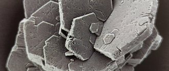
Stones that form in the urinary organs have a varied toxic composition of chemical elements. Quite a lot of them can form. They are also completely different in shape.
Urethrostomy surgery for urolithiasis in males and females
Urethrostomy surgery for urolithiasis in males and females
Konstantinovsky Alexander Andreevich
Veterinary surgeon.
Veterinary ophthalmology specialist. Diploma winner from the Ministry of Education of the Russian Federation. Area of scientific interests: ophthalmology, eye microsurgery, plastic, vascular, optical reconstructive surgery.
Chief physician of Doctor Meow LLC since January 2007
Urolithiasis (urolithiasis)
Urolithiasis is a disease involving the formation of urinary stones in the kidneys or urinary tract. The formation of stones can be a consequence or cause of chronic inflammation of the urinary tract. The incidence of urolithiasis varies between breeds and regions, ranging from 0.4 to 6%. It depends on genetic, metabolic, environmental and nutritional factors.
Bladder stones are the most common urinary calculi. They irritate the mucous membrane and, when released through the urethra, lead to blockage of the urinary tract in males and female cats. Type and relative frequency: stones containing magnesium and ammonium phosphate (70-90% of all stones); stones containing calcium oxalate (5-8%); stones containing calcium phosphate (1-3%); urate stones (2-4%, very envious of the rock); cystine stones (2-22%, depending on the breed, 95% of cases in males); silicate stones (1-3%) and mixed stones (5-10%).
Symptoms
Symptoms vary depending on location and gender. Blockages of the urethra are found mainly in males and female cats (long, curved narrow urethra, penis bone in males). In this case, there is a partial inability to urinate, painful, drop-like urination (stranguria) or an enlargement of the bladder and abdominal cavity with complete blockage. If the urethral blockage is not cleared, postrenal renal failure develops within 48 hours. Bladder ruptures due to urolithiasis are rare.
Females do not experience blockage or pollakiuria due to the short, wide urethra. Hematuria in the initial or final portion of urine, stranguria, dysuria, pain on palpation and, possibly, thickening of the walls of the bladder come to the fore. Bladder stones can go undetected for a long time and are discovered by chance. Tripelphosphate stones in breeds with a predisposition to urolithiasis (schnauzers) are observed at an early age. In addition, a predisposition to the formation of stones is characteristic of male dachshunds and basset hounds (cystine stones), Dalmatians (urate stones). In cats, any breed predisposition has not been identified, however, in castrated cats, the incidence of urolithiasis is slightly more common, although this does not give the right to talk about the tendency of castrated cats to urolithiasis.
Analysis of urine
Microhematuria up to the most severe hematuria, signs of inflammation; often, although not always, alkaline pH and crystallurgy.
Diagnosis verification
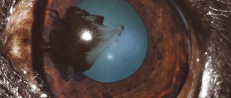
A plain film of the bladder and urethra is the main means of detecting stones that are opaque to x-rays (phosphates, calcium oxalate). In unclear cases and with low mineralization of stones (urate), a cystogram of the urethra using positive contrast agents or a double-contrast radiograph can be done (air bubbles may look like stones; they are hexagonal in shape and stay near the mucous membrane, while the stones are visible mainly in the center of the bladder). Large stones can be palpated. Stones in the urethra can be detected using a catheter or probe through the penile bone or sciatic notch. A good diagnostic method is ultrasound diagnostics, which in most cases makes it possible to accurately diagnose not only bulky stones in the bladder, but also small suspensions that can injure the urethra.
Predisposition and causes
Predisposition and causes are not fully understood. Reliable factors are:
- Oversaturation of urine with minerals under certain pH conditions contributes to the precipitation of crystalline substances.
- Deficiency of factors that stabilize the composition of urine, the presence of foci of crystallization (incorrectly sutured bladder during cystotomy).
- Endogenous or caused by increased intestinal absorption, an increase in crystalloid loss.
- In situ formation of phosphates or urates enhanced by bacterial activity.
- Stagnation of urine, long intervals between emptying the bladder.
- Genetically determined metabolic abnormalities (cystine stones, urate stones). The effect of other factors, such as vitamin A deficiency, is unclear.
I distinguish between infection stones (phosphate stones) and metabolic stones (cystine, urate, silicate and oxalate).
Complications
Nephrolithiasis (formation of kidney stones), urethritis and pyelonephritis, hydronephrosis, azotemia, chronic hypertrophic cystitis (bladder with severe muscle hypertrophy) with chronic partial blockage of the urethra by urinary stones, renal failure.
Forecast
Except in complicated cases, favorable. Without prevention of stone formation, relapses are inevitable.
Treatment
A distinction must be made between emergency measures, surgical or conservative stone removal, and stone prevention.
Emergency measures
Depending on the general condition, a blood sample is first taken and the presence of azotemia and renal failure is established, or the urethral blockage and bladder emptying are immediately started. For this you can use:
- retropulsion method;
- insertion of a thin catheter to empty the bladder;
- crushing stones using an ultrasonic device;
- cystocentesis.
- Retropulsion method: return of stones to the bladder, from where they can be removed by cystotomy or dissolved by conservative methods. a) Insert the urinary catheter until the stone is reached. b) Inject a lubricant diluted in a 1:1 ratio to reduce friction, possibly introducing a local anesthetic (2% lidocaine). c) Resisting animals should be administered an analgesic, antispasmodic, or short-acting narcotic such as a thiobarbiturate or inhalational anesthetic. d) Insert a suitable nipple cannula or catheter into the penis. Connect a syringe containing 20-60 ml of warm, sterile sodium chloride solution to the cannula or catheter. With your index finger, squeeze the urethra per rectum, inject sodium chloride solution until the urethra is distended, then either in the distal part, quickly remove the nipple cannula or catheter, as a result of which the stone should move towards the urethra, or in the proximal part, releasing index finger, release the compression, as a result of which the stone should be washed away towards the bladder. A Foley catheter is better than a nipple cannula for flushing stones into the bladder. e) Repeat until the stone gets back into the bladder (in about 80-90% of cases this is possible). Empty your bladder and later promptly remove the stones.
- Emptying the bladder. A thin, well-lubricated urinary catheter through which a sodium chloride solution is injected under pressure can sometimes be passed past the stone and into the bladder. This way you can empty your bladder.
- Stone crushing using a special probe connected to an ultrasonic device for subsequent removal of stone fragments.
- Cystocentesis (bladder puncture) is used as a last resort, when it is not possible to quickly remove the blockage of the urethra, and the animal should be prepared for long-term surgical intervention.
Surgery
Surgical treatment of urolithiasis involves removing stones from the bladder and creating a urethrostomy in a wide area of the urethra, or above the stone or stricture.
Removing bladder stones (cystotomy) is the main method for removing stones that cannot be dissolved. The operation should not be performed only if the risk associated with anesthesia is disproportionately high, or if the owner is against the operation. In these cases, conservative treatment can be carried out in combination with the creation of a permanent urethral fistula (urethrostomy).
Operating technique - cystotomy
Laparotomy is performed in females and males along the linea alba or along the midline in males. During surgery, a permanent catheter should be inserted to drain urine and to move stones into the urethra. Stretch the bladder using threads and isolate it from the abdominal cavity before opening. Remove stones with a spoon. Wash the sand. Before double-layer bladder closure, carefully check for stones in the bladder neck. Compare the number of stones removed with the number of stones visible on the x-ray, and in doubtful cases, carry out radiographic control.
Operative technique - urethrostomy
Depending on the specific case, three types of urethrostomy are performed on male dogs: distal, scrotal and perineal. In cats, perinenal urethrostomy with amputation of the penis is performed.
Distal urethrostomy (in dogs)
Indications
The presence of a calculus in the urethra at the caudal end of the bone of the penis. It is worth noting that in most cases the stones are localized in this location.
Technique
The outer covering is pulled caudal to the bone of the penis and raised along with the penis. Typically, a stone or the button end of the probe is palpated through the skin. The incision is made along the suture above the blockage site exactly along the midline, while periodically monitoring the position of the stone or probe head by palpation. At the same time, I cut the skin by 2-3 cm, and also after dissecting the paired muscle that pulls the penis back - the corpus spongiosum of the penis and the mucous membrane. Urethral stones located caudal to the incision site often flush out spontaneously under the pressure of a full bladder. If necessary, they are removed with a loop catheter. Stones located cranially are squeezed out using a probe. Firmly seated stones can be removed using a small spoon corresponding to the lumen of the urethra.
By inserting an elastic urethral catheter and rinsing with a cold electrolyte solution to the bladder, the patency of the urethra is checked.
If a suture is not applied, healing of the urethral incision usually occurs without stenosis within several weeks. However, to prevent significant bleeding from the cavernous (cavernous) body and subcutaneous infiltration of urine, it is recommended to temporarily sew the edge of the mucous membrane of the urethra to the edge of the skin wound with thin atraumatic threads like a circular suture.
During urethrostomy, in order to create a permanent fistula, the threads are left until the scarring is complete.
Sometimes in the future there is a need to close an artificial external fistula of the urethra. To do this, the urethra is separated from the skin as evenly as possible. It is important that when grasping the urethral wall, the tissue is not damaged. The free edge is sutured over the inserted urethral catheter with absorbable atraumatic threads (1-2 thick) like an interrupted suture. At the same time, try not to puncture the mucous membrane. Then the skin wound is sutured.
A catheter (preferably hard) is fixed to the abdominal wall to ensure urination for several days.
Scrotal urethrostomy (in dogs)
Indications
Stricture of the distal part of the urethra, urolithiasis.
Technique
After castration, the scrotal skin is excised in the shape of an ellipse around the suture of the scrotum, so that the remaining part of the incision edge can be aligned without tension and the formation of a pouch-like expansion. In the caudal part of the wound, the urethra is opened bluntly and an incision about 3 cm long is made on it. The edge of the mucous membrane is sutured to the skin with atraumatic non-absorbable suture material. In the cranial part, the edges of the skin are sutured.
Perineal urethrostomy (in dogs)
Indications
Stones in the urethra in the perineal area.
Technique
The anus is closed with a purse string suture or a tampon sewn on top. The skin is incised along the midline at the level of the ischial notch. In this case, you can palpate the urethra, isolated using a catheter. Access to the urethra is opened by blunt preparation between the paired muscle that pulls the penis back and the same paired bulbospongiosus muscle. After this, I make an incision in the urethra about 3 cm long, and then the stones are removed by washing or using a spoon or loop catheter. The urethral catheter is inserted up to the bladder.
The edges of the mucosal incision are sutured to the edges of the skin wound with atraumatic non-absorbable threads.
Perineal urethrostomy with amputation of the penis (in cats)
Indications
Blockage of the urethra.
Technique
The skin is cut in a circle around the opening of the foreskin. An incision is made along the suture of the scrotum to a point located at a distance of about 15 mm ventral to the anus. Then, if the cat is not castrated, castration is performed with the testes closed and the spermatic cord closed. After this, the penis is exposed to the area located proximal to the bulbourethral glands. First, the ischiocavernosus muscle is dissected on both sides along with the ischiourethral muscle and the bleeding from the blood vessel on the muscle stump is stopped. The muscle that pulls the penis back is then incised, just distal to the ventral loop of the rectum and also close to the glans, and the urethra is cut above the catheter. Using scissors, the holes are widened to approximately 25 mm to a place located proximal to the bulbourethral glands, and one mosquito clamp is applied to the edges of the urethral incision on both sides.
If before this it was impossible to insert the catheter, now, after removing the stone with a small blunt spoon, it is advanced to the bladder. In this case, it is necessary to carefully ensure that urine does not flow into the wound. The edges of the urethral incision are then sutured to the edge of the skin wound (slowly absorbable synthetic thread).
The first stitch is made in the proximal corner of the wound. The needle is passed through the skin, at an angle to the edge of the incision and perpendicularly, through the wall of the urethra from the outside to the inside, and on the other hand - from the inside to the outside through the wall of the urethra, and also at an angle through the skin in the opposite direction. Then the wall of the urethra is sutured with an interrupted interrupted suture to the skin along the length of the incision. To avoid uneven tension on the edges of the wound, stitches are applied alternately on one side and the other.
At the peripheral edge of the urethral incision, the penis is ligated (absorbable sutures) and separated.
Then reverse stitches are applied to the proximal corner of the urethral wound. Wound Closure
The wound edges of the superficial fascia and subcutaneous layer are sutured over the stump of the penis using interrupted stitches (absorbable suture material). Then a skin suture is applied.
Follow-up treatment and prevention of urinary stone formation
Antimicrobial treatment of cystitis as long as stones are present.
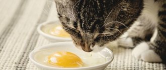
Reduce triggers, such as oversaturation of urine with stone-forming crystalloids, by increasing water intake and reducing the intake of minerals from food. To do this, give liquid food 3 times a day with the addition of 1-10 g of salt per day for an unlimited time (it is advisable to achieve a urine specific gravity of 1.020). If cystine and oxalate stones are present, salt should not be given. The animal should always have fresh water in unlimited quantities.
Specific measures (depending on the type of stones). To do this, you need to examine urinary stones or urinary sediment. Chemical analysis of urinary stones is easy and inexpensive, but does not provide very accurate information. In all cases of recurrence or complications, it is recommended to refer patients to the more expensive X-ray diffraction or infrared spectroscopy examination. If there are no samples of stones for examination, you can try to identify typical crystals in the urinary sediment or treat as for phosphate stones, since this type of stones occurs in 90-95% of cases in all dogs except Dalmatians and breeds with a predisposition to the formation of cystine stones.
After determining the nature of the stones, you can choose a special diet for your pet, which will help dissolve existing stones and prevent the appearance of new ones. The mechanism of action of diet therapy is simple: certain foods can change the pH level of urine, and for the formation of stones a certain level of acidity or alkalinity is required (depending on the nature of the stones). There are a variety of premium and super premium therapeutic diets on sale, created for the prevention and treatment of urolithiasis. You can check the availability of these diets at your nearest pet store.
Literature:
1. H.G. Niemand, P.B. Suter “Diseases of dogs”, Moscow, “Aquarium”, 2001 2. H. Shebits, V. Brass “Operative surgery of dogs and cats”, Moscow, “Aquarium”, 2001 3. A. Kuzmin “Aibolit’s advice or the health of your dog” , Kharkov, “Paritet”, 1996 4. F. Barr “Ultrasound diagnostics of dogs and cats”, Moscow, “Aquarium”, 2001 5. Michelle Bambrger “The quick guide to first aid for your cat”, New York, 1995
Why is it dangerous?
If your cat has a large blockage, then as a result, his bladder begins to stretch. As a result of its stretching, all the blood vessels on the walls of the bladder burst. In this case, the blood further enters the animal’s urine. Or vice versa: urine into blood. Due to this, the body is poisoned by various residual chemicals in the urine that should have left it.
As a result of the above processes, pressure in the bladder increases . It spreads higher to the kidneys, affecting them so that they become tense and then hemorrhages occur. Obstruction greatly disrupts the normal functioning of the kidneys, reduces the level of blood circulation in them and negatively affects filtration processes. Urinary retention in the body leads to the development of azotemia. This is a pathology that consists of an elevated level of nitrogenous substances in the blood.
© shutterstock
Through this pathology, uremia develops further. This is self-poisoning of the body, which occurs due to severe impairment of kidney function. If timely surgery is not performed on a cat with urolithiasis, the bladder may rupture.
How is surgery performed?
Planned surgery is carried out only after anesthesia. The animal is fixed on the operating table in the position “lying, on its back, with the pelvic limbs extended backward.”
At the beginning of the operation, a skin incision is made in the shape of an ellipse in the area of the foreskin and scrotum. The excised cat genitals are separated from each other and removed. The penis is also separated from the fatty tissue and removed.
Afterwards, the ischiocavernous and ischiourethral muscles are alternately dissected. If there is heavy bleeding from a blood vessel, measures are taken to stop the bleeding. Next, the muscle that pulls the cat's penis back is cut, and the urethra, which is located above the catheter, is cut. Then the stitches are made.
The first suture is placed in the proximal corner of the wound from the outside in and from the inside out using a special technology. The urethral stitches are sutured to the skin along the entire length of the incision. When applying sutures, much attention is paid to uniform tension of the wound edges.
The penis is then coagulated or bandaged and separated. Reverse sutures are placed at the proximal angle of the urethral wound. As a result of the operation, a formed stoma is formed in the wide part of the urethra and bougienage is performed.
Why is the disease common in cats?
It is believed that this disease is observed in cats as often as in cats. But no one usually notices her. After all, obvious manifestations are not typical for her. This also applies to dogs and cables. All this is connected only with anatomy. In cats, the urethra, that is, the urethra, is structured two times shorter and wider than that of a cat. This allows small stones and sand to naturally pass out of the body through urine. Unlike cats, a cat's urethra is long and narrow, so when sand wants to come out, it forms a blockage.
The urethra always gets clogged in the part that is the narrowest. But if the stones are large in size, then a blockage can form near the bladder itself.
In order to restore normal urine flow in a cat, veterinarians need:
- remove the blockage of the canal using a catheter (or urethrostomy);
- stabilize the general condition of the cat.
How to prepare a cat for surgical treatment?
Urethrostomy in a cat is carried out only after a complete examination of the animal, clinical studies and laboratory tests.
In particular, a few days before surgical treatment, the following is carried out:
- ultrasound examination of the abdominal cavity;
- radiography of the urinary system;
- general urine analysis;
- biochemical and general blood test.
An uncastrated cat undergoes mandatory castration. Before surgery, the animal's urethra is washed and the bladder is catheterized, from which stones and sand are removed. Before surgical treatment, it is strictly forbidden to give your pet food to avoid complications.
Subtleties of the operating process
If obstruction (blockage) of the urethra in a pet does not disappear with the help of conventional medicinal methods and relapses occur again, surgical intervention should be resorted to, that is, a urethrostomy is performed in the cat.
So, urethrostomy is a surgical type operation. It lies in the fact that a new urethral formation is formed following the example of the feminine gender (short but wide).
There is such a division:
- perineal urethrostomy in cats;
- prepubic urethrostomy;
Pre-pubic urethrostomy is used by veterinarians to correct various types of pathologies that have formed in the pelvic area. The method is considered a last resort when the perineum cannot conduct urine.
Typically, doctors choose perineal urethrostomy. In its course, a new creation is formed. During this operation, if the cat is not neutered, he is castrated. As a result, the leading channel becomes wide and straight, so sand and small stones can easily pass through it.
Urethrostomy of a pet is done so that in the future obstruction, that is, blockage, does not occur again. But we should not forget that with its help, the cat’s urolithiasis does not disappear anywhere. She requires further treatment. However, this is not surgical, but only therapeutic.
Types of urethrostomy
- perineal - the most common method, performed to prevent blockage of the urethra. Surgical access is performed in the area of the cat's penis and scrotum, the surgeon excises the genital organs (penis and testes) and begins to form a stoma by suturing a wide, straight part of the urethra to the skin of the anterior abdominal wall or perineum, depending on the surgical technique used;
- prepubic - this method is more complicated, it is used in cases of inaccessibility of the first method: with traumatic ruptures and neoplasms of the urethra, incorrect perineal urethrostomy.
Indications
Urethrostomy in a cat requires good reasons to do it.
Indications for urethrostomy:
- repeated occurrence of relapses of urolithiasis;
- obstruction;
- various, but only significant disorders and damage to the genital organ in cats;
- urinary retention.
- various types of deformation of the channel through which urine passes;
- if catheterization of the bladder is not possible.
In order to perform a urethrostomy, doctors must clearly identify in advance the place that is clogged with stones. Because this particular operation is done in order to remove the two narrowest sections of the urethra. After all, it is in them that sand and various large formations most often collect.
Why do plugs form and urethrostomy is required?
There are several reasons why a plug forms in the urethra of cats. Most often this is a symptom of urolithiasis - in this case, the plug is formed by salts. Often, blockage of the urethra occurs with cystitis, and the plug is formed by clotted blood cells. Traffic jams can form due to injuries to the penis and inflammatory processes. In rare cases, the urethra becomes blocked due to tumors or diseases of the nervous system that cause spasm. Canal obstruction may result from previous surgical procedures. In each case, the cat should undergo a urethrostomy as soon as possible to avoid possible complications.
What about after the operation?
In order for the swelling to disappear after urethrostomy, and for the newly formed opening to remain as wide as it was made, bougienage is made during postoperative rehabilitation. This procedure is instrumental and non-surgical. It involves inserting a wide probe into the urethra, or it can be a catheter. It is used to check whether the urethra is sufficiently open.
The veterinarian carefully cleans the stitches. At the same time, they must definitely check the correctness of the hole made during urethrostomy. It is worth watching to ensure that the cat does not scratch or lick the wound . Therefore, you definitely need to purchase a diaper and a protective collar. This way the wound will be inaccessible to the animal, and it will heal calmly. To prevent inflammation from forming on the wounds and causing them to fester after urethrostomy, and to prevent bacterial cystitis or urethritis from developing, veterinarians prescribe medications that have an antibiotic effect.
© shutterstock
To ensure that the damage does not spread to the kidneys, the veterinarian monitors the general condition of the cat.:
- whether he drinks enough clean and purified water;
- does he go to the toilet after that?
- what is the cat's appetite?
- or it is active.
Complications of urethrostomy
The most common early complication of this operation is bleeding at the sites of tissue dissection. No treatment is required; the bleeding goes away on its own.
The most common late complication is a sticture, which is a consequence of an incorrectly performed operation, a long stay of the catheter in the urethra and self-injury of the animal.
A bacterial infection may develop due to the loss of natural defense mechanisms against microorganisms, which are provided by narrowing of the urethra. Sometimes this leads to the spread of an ascending infection in the urethra. Most often, infections are provoked by manipulations during treatment (for example, catheterization) and relapses of urethral obstruction.
Urethrostomy should be performed by an experienced veterinarian. After the operation, measures should be taken to prevent self-injury to the animal, such as a postoperative blanket and collar.
(c) Veterinary center for the treatment and rehabilitation of animals “Zoostatus”. Varshavskoe highway, 125 building 1. tel.
8 (499) 372-27-37
Symptoms
If you notice a decrease in your cat’s temperature after urethrostomy, he categorically refuses to eat, his muscles twitch, then this could be either uremia or azotemia. In this case, you should immediately seek help from a veterinarian!
After 10-14 days, when the urethrostomy was performed, and when the cat was fully restored after the urethrostomy, the sutures are removed. But this is done only after the clinic worker is completely convinced that they have healed and there is no inflammation on them.
They also pay special attention to whether the newly formed stoma is functioning normally. Animals who have undergone urethrostomy need to be examined and tested every six months.
Urethrostomy in a cat consequences
Within a few days after surgery, minor bleeding may appear, which does not require additional treatment. In operated animals, there is a high probability of secondary infection of the formed canal.
In rare cases, urinary incontinence may develop. Most often, a cat recovers after urethrostomy surgery after a short time (2-4 weeks) and, with proper care, continues its usual lifestyle. If the hole is noticeably overgrown with scar tissue, repeated intervention is performed. Stricture can be prevented by bougienage in the postoperative period and antiseptic treatments.
Should we expect any problems?
As always, after every surgery something can go wrong. Therefore, there may be complications after urethrostomy in a cat. Let's look at each of them in more detail. Urethrostomy accompanied by difficulties is normal.
Bleeding. This complication is not life-threatening. Because of this, there is no need to perform a urethrostomy on the cat again. You can also independently determine the degree of its severity using the color of the mucous membrane. If it is still severe and serious, then it can be removed under general or local anesthesia.
Anuria may also occur. It is characterized by the fact that urine stops flowing into the cat’s bladder. Therefore, the cat may not go to the toilet for approximately two days. But after urethrostomy, such a violation is very rare.
One of the complications can be acute kidney failure. It can occur if the veterinarian did not notice that the cat had pathologically enlarged kidneys before the urethrostomy. Such a disorder can be detected after tests for urethrostomy have been taken. It can be eliminated with the help of appropriately prescribed treatment.
© shutterstock
How to care for a cat after surgery
Caring for a cat after surgical treatment involves following certain recommendations of a veterinary surgeon. If the procedure is successful, hormonal and antibacterial therapy is prescribed, as well as local treatment with ointments and dietary nutrition.
In case of successful operations, the sutures are removed on the 12th day.
In case of swelling, swelling, redness, suture separation and other problems, the sutures can be removed later:
- Antibacterial therapy. To avoid consequences and repeated urethrostomy, antibiotics are prescribed. The dosage is determined in each case individually, taking into account the age and weight of the animal. The rehabilitation course lasts on average up to 7 days.
- Hormonal therapy. After surgery, cats are prescribed hormonal medications to relieve swelling. The most effective are dexamethasone or prednisolone in the form of injections. The course of recovery with hormonal agents takes up to 3 days, depending on the condition of the sick animal.
- Local treatment with ointments. In the first days after bougienage, an ointment (for example, mastitis forte) is applied to the operated area for 1-2 days. In the following days, a 0.05% solution of chlorhexidine bigluconate and mucosanine is applied to the problem area. Seams are processed daily, 2 times a day. The course of local treatment lasts on average up to 2 weeks.
- Diet food. To restore strength, it is important for operated pets to eat properly and limit movement. It is necessary to choose the right food and not burden the cat’s body.
If your pet has various pathologies of the urinary system that cannot be treated with medication, surgery is simply inevitable. Timely urethrostomy in a veterinary clinic will help maintain excellent health of the cat for many years.
After treatment
When a little time passes after urethrostomy in a cat, the formed urethra may suddenly begin to narrow, that is, the processes of overgrowth begin. Here you will need to do a urethrostomy again. The fact that the urethra begins to overgrow may be a consequence of the operation itself being performed incorrectly by veterinarians, or it could be torn somewhere due to certain injuries. This complication is rightfully considered the most serious and severe.
Another complication that urethrostomy brings is often dysuria. In general, this disease disrupts the cat’s urination process. The underlying cause here may be the presence of certain bacteria in the urine after urethrostomy. Or it may be due to the costs of urological syndrome. Another reason leading to this complication is stones or a tumor located in the bladder. As already mentioned, urethrostomy does not cure urolithiasis, but only removes blockages.
Postoperative care
In order to prevent overgrowth of the stoma, a urinary catheter is sewn into the animal for 1-3 days. Drug therapy is prescribed, including the use of antibacterial and painkillers, as well as local treatment of sutures with antiseptic solutions. To prevent stitches from licking, the animal is wearing a protective collar, which must be worn for 7-14 days.
After removal of the urinary catheter, the stoma is subject to regular bougienage (insertion of a soft urethral catheter) - a procedure that allows you to check the patency of the stoma and urethra, as well as prevent them from becoming overgrown.
The sutures are removed from the skin on the 10-14th day.
Since in case of recurrent idiopathic cystitis and urolithiasis, urethrostomy is only a symptomatic treatment of obstruction, after the operation it is necessary to continue therapy for these diseases (the use of medicinal feed, diuretics and sedatives).