Nature has wisely taken care of reliable eye protection with the help of eyelids, eyelashes and tear fluid, but they do not save you from injuries due to negligence. Due to various reasons, a scratch on the cornea of the eye and the appearance of unpleasant symptoms accompanying it are quite common occurrences. Self-medication in such cases is unacceptable; you should consult an ophthalmologist as soon as possible. Only a specialist can accurately determine the extent of damage, professionally provide first aid and prescribe treatment if necessary.
How to tell if your cornea is scratched
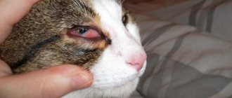
The cause of damage to the delicate cornea can be dust or a foreign body, an impact, prolonged wearing of contact lenses or the presence of chips on them, intense friction when a speck gets in. Sometimes the first signs of a scratch on the cornea do not appear immediately, so it can be difficult to determine the source of the problem. But characteristic symptoms convincingly indicate the presence of injury:
- pain that intensifies when trying to open the eye;
- severe lacrimation, redness;
- sensation of a foreign body in the eye;
- photophobia, lack of clarity of visual perception;
- headaches often occur.
If possible, you should immediately consult a doctor, but first try to alleviate your condition.
The animal needs to be calmed down
Remember that the best sedative is kind, comforting words spoken by the owner in a gentle and calm tone. Panic and screaming, on the contrary, will make the cat even more anxious. In addition, you can give your pet a little sedative, for example, “Cat Bayun”. When the animal has calmed down, you can wash the fresh scratches with a solution of hydrogen peroxide and wipe the wounds with a gauze pad that needs to be soaked in brilliant green. Algorithm for treating wounds.
In case of any, even the most minor injury, the wound still needs to be treated with antimicrobial drugs. We propose to do this using the following algorithm. After the bleeding has stopped, carefully trim the hair that surrounds the damaged area. If there are foreign objects in it (sand, grass, pieces of glass), they must be removed with tweezers. Next, we treat the wound with three percent peroxide or a weak solution of potassium permanganate in order to eliminate bacterial flora and remaining dirt. We recommend disinfecting the skin around the wound with a solution of brilliant green, alcohol or iodine.
This is followed by treatment of the damage with “Sanatol” or “Streptocide”. If you see a deep defect, then you first need to stop the bleeding, treat everything according to the above method and then apply a sterile bandage to the wound, the edges of which are fixed with a regular adhesive plaster.
First aid
If a small speck or eyelash gets into your eye, you need to blink frequently - increased tear flow can sometimes remove the disturbing small object. If there is no result, you should connect the eyelashes - carefully pull the upper eyelid over the lower eyelid. If this measure does not help, you should try to rinse the eye - always only with clean water or physiological (saline) solution with a comfortable temperature of 20-37°C. To do this, you need to lower your eye into the container with the solution and blink vigorously. It’s a good idea to drip drops that moisturize the mucous membrane - they are available in every pharmacy without a prescription. Even if first aid was effective, it is still worth making an appointment with an ophthalmologist for an examination. Removal of a foreign body does not mean the absence of a scratch on the cornea of the eye - how long the injury heals will depend on the timeliness of the prescribed treatment. And do not forget about caution - all of the above measures are not applicable in severe cases, when an urgent visit to an ophthalmologist is necessary.
Eye loss
Another common injury is eye prolapse. Any animal can receive such damage, but do not forget about breed predisposition.
Traditionally, the risk group includes Pekingese, Pugs, Chihuahuas, Spaniels, Boxers and English Bulldogs. These dogs are traditionally distinguished by a short muzzle, flattened head shape and large, protruding eyes. If your pet's eye falls out, you should immediately go to the veterinary clinic. It is absolutely forbidden to correct a prolapse on your own!
It is very important that the eye does not dry out during the journey. Therefore, as an emergency measure, it may be advisable to coat the damaged visual organ with gel. And under no circumstances should there be any bandages! Contact of a bandage, like any other sterile material, with the surface of the eye in such a situation is fatal!
How long does it take for healing to occur?
It is difficult to definitively answer the question of how long it takes for a scratch on the cornea of the eye to heal. When it is possible to get rid of a foreign body by washing or tear drops, a few days are enough. After an eyelash gets into the eye, the unpleasant symptoms gradually disappear over 1-2 days. But sometimes an infected scratch or excessively deep damage requires adequate treatment; this can only be determined by a specialist. Using an ophthalmoscope, the doctor determines the degree of damage and removes the foreign body if present. Depending on the result of the examination, the optimal treatment for a scratch on the cornea of the eye is selected:
- antibiotic ointment;
- antibacterial drops;
- steroid drops to eliminate inflammation.
During treatment, it is recommended not to use cosmetics and contact lenses. Sunglasses help protect your eyes from bright light. If you follow your doctor's recommendations, a scratch on the cornea of the eye will heal in about a week. However, sometimes, if a complication occurs or the damage is deep, longer complex treatment is required.
What eye injuries can a cat have?
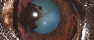
Bruising in a pet's eye can occur as a result of exposure to sharp or blunt objects. If the damage was caused by blunt objects, then they are safer, because in this case the eyeball does not suffer. If the wound is caused by sharp objects, then the following may occur:
- displacement of the eye lens;
- severe opacities in the anterior chamber of the eye;
- damage to bone tissue near the eye socket;
- detachment of the eye cornea;
- profuse bleeding.
Most often, symptoms of eye injury are observed in the spring, when cats go into labor. In addition, such damage is often observed in pets that live near major highways. Small stones can jump out from under the wheels of passing cars and harm the animal.
However, the damage is not in all cases mechanical in nature. Sometimes injuries occur due to excessive care of the owner, who gives his pet strong veterinary drugs without first consulting a specialist. For example, unnecessary or inappropriate antibiotic therapy can cause complete loss of vision or hearing. If your pet's condition worsens, it should be shown to a veterinarian as soon as possible.
How to protect yourself from corneal injury
As a rule, a scratch on the cornea of the eye appears under the influence of external factors, and often the injury could have been avoided. When performing work that increases the risk of injury, you should wear safety glasses. This applies to operating power tools and chemicals, cleaning premises and mowing lawns, playing certain sports, riding a motorcycle, and performing metalworking work.
Scratches on the cornea can be caused by careless use of mascara or prolonged wearing of contact lenses. They cause excessive dryness of the cornea, making it more vulnerable. Wearing poorly adapted or damaged lenses is especially dangerous. Dry cornea increases the risk of injury; using moisturizing drops will help eliminate the problem.
Possible consequences
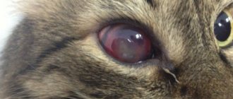
Complications that may occur after hemorrhage depend on the degree of filling of the anterior chamber. If there is an outpouring of blood on the area of the cornea, its color changes, and accordingly, the quality of vision decreases. Even after the blood is removed, such a defect can remain for a long time until it gradually disappears. Due to a sharp increase in IOP at the time of injury, secondary glaucoma can also develop. Moreover, the greater the volume of blood shed, the higher the risk of its appearance and decreased vision.
Sometimes anterior synechia also occurs - adherence of the iris to the cornea. In combination with increased IOP, this can provoke death of the optic nerve. The greater the amount of blood in the eye, the worse the quality of vision becomes.
What are the consequences of not visiting a doctor?
As a rule, vision loss is rarely caused by a simple scratch to the cornea of the eye. But sometimes its consequences are quite serious. First of all, trauma can negatively affect the exacerbation of existing eye diseases. The consequences of a late visit to an ophthalmologist can be:
- inflammation;
- formation of purulent exudate;
- spread of infection deep into the eye, sepsis;
- corneal edema, scarring, pupil displacement;
- increased intraocular pressure;
- traumatic cataract, which is accompanied by clouding of the lens and a gradual decrease in vision;
- brain abscess.
People with weak immunity and various metabolic disorders are at risk. But even for healthy people, a scratch on the cornea can cause serious consequences. Correctly provided primary care is of great importance, so visiting an ophthalmologist is extremely important.
Clean View Clinic specialists provide high-quality diagnostic and treatment services for all types of eye diseases.
The center's website contains up-to-date information; you can make an appointment online or by phone. High-quality equipment and many years of experience of the best ophthalmologists in Moscow will help determine the severity of the injury, prescribe the correct treatment and avoid further vision problems. At the Clean View clinic you can undergo a full range of procedures for diagnosing vision and treating eye diseases. More details by phone.
Hemorrhage into the vitreous body of the eye: symptoms and treatment
A blow to the vitreous area results in hemorrhage. Blood in the retrolental space of the vitreous expands it, and blood in the orbicular space leads to the formation of a specific rim (stripe) that surrounds the periphery of the lens behind.
Retrolental hemorrhage takes longer to resolve than orbicular hemorrhage. Sometimes minor hemorrhages may be invisible and are discovered later, when they “descend” to the lower part of the anterior chamber.
Hemophthalmos is understood as massive hemorrhage into the vitreous body, occupying a significant part of the latter.
Approximately on the third day after injury, the blood in the vitreous body undergoes a process of hemolysis with the loss of hemoglobin by red blood cells, as a result of which they become colorless and later disappear. And the hemoglobin of erythrocytes takes on the form of grains, which are subsequently absorbed by phagocytes. Hemosiderin is formed, which has a toxic effect on the retina. Sometimes the blood does not completely resolve and a blood clot is formed and replaced by connective tissue moorings. The clinical picture of hemophthalmos is dominated by loss of visual acuity from the state of light perception to complete blindness. Focal illumination and biomicroscopy make it possible to see behind the lens a dark brown granular, sometimes with a reddish tint, mass of blood that permeates the vitreous body. Ophthalmoscopy shows the absence of a fundus reflex. Later, when the blood clot dissolves, one can observe deformation of the vitreous with its liquefaction. Hemophthalmos must be distinguished from partial hemorrhage into the vitreous body, which quickly and completely resolves.
Hemophthalmos leads to the development of degenerative processes in the vitreous body.
Treatment
In case of vitreous hemorrhage, bed rest and a cold bandage are prescribed on the affected eye. Calcium preparations (tablets, eye drops and intramuscular injections), hemostatic agents (Vicasol) are used. To speed up the resorption of hemorrhage, heparin is used (subconjunctivally on days 1–2) and enzyme preparations and potassium iodide. Treatment of hemophthalmia: bed rest with the head end elevated. Use a binocular bandage for 2–3 days. Calcium chloride, pilocarpine 1% 2 times a day, glucose with ascorbic acid are used, and a solution of dicinone (12.5%) is injected subconjunctivally. Then, after 2-3 days, absorbable drugs are used: dionin, potassium iodide, lidase. Corticosteroids (under the conjunctiva) and fibrinolysin are also indicated. In the late period of treatment, ultrasound and physiotherapy help a lot. If there is no positive effect from therapy, then it is necessary to suction the vitreous, and sometimes excise part of it. The removed vitreous body is replaced with luronite (a hyaluronic acid preparation).
We must not forget that with somatic diseases (cardiovascular diseases, atherosclerosis, hypertension, blood diseases, endocrine pathology), the development of hemophthalmos is possible. But in these diseases, hemophthalmos occupies a small part of the vitreous body.
The prognosis depends on the area of hemorrhage. If the hemorrhage covers 1/8 of the vitreous body, it often resolves. With an area of 1/8-1/4 of the vitreous, moorings are formed, which leads to retinal detachment. In terms of restoring visual functions and preserving the organ in total hemophthalmia, when a blood clot occupies more than ¾ of the vitreous, the prognosis is unfavorable. Irreversible destructive changes occur in the vitreous body (organization of a blood clot, formation of adhesions). Tractional retinal detachment and atrophy of the eyeball may develop.