Dogs quite often suffer eye injuries due to an accidental blow while running, a fight with other animals, a fall from a great height, or a car accident. The consequences of this may be irreversible. At best, traumatic damage to the organ of vision can lead to irritation, and in the worst case, to blindness and loss of the eye.
Timely assistance will prevent corneal destruction and maintain excellent vision for many years.
When to seek veterinary help?
An eye injury in a dog is a serious problem that requires immediate attention to a veterinarian. There are various problematic situations in which it is necessary to rush to the veterinarian.
- Getting injured as a result of an unpleasant interaction with a street cat. A fight between an angry cat and a dog can be costly for the animal's health. If in the first hours after the injury your pet rubs its eye vigorously, squints and rushes from side to side in pain, it is necessary to carefully examine it. If, after an injury, clouding of the cornea in the area of injury, changes in the eyeball, or the appearance of blood or pus are clearly visible, you should immediately go to a veterinary clinic, where a specialist will prescribe the appropriate treatment.
- Mechanical damage due to debris, dust, or sand getting into the eye. When a small foreign body penetrates under the third eyelid, severe irritation appears on the cornea of the eye. The dog will try to scratch his eye with his paw and squint all the time. He will experience discomfort and pain. If excessive tearing occurs, the dog owner needs to be wary. In this case, it is necessary to remove a speck of dust or speck from the eye in a veterinary clinic. Otherwise, complications may occur, including blindness.
- Traumatic injury due to penetration of the eye by a sharp object. When a branch, stick or other object gets into the eye, the cornea is damaged and a wound is formed into which various microbes and bacteria penetrate. If the dog has an urgent need to squint, this may also indicate a minor injury. With a strong blow, an injured animal may completely close its eyes and experience pronounced painful sensations. If at the same time purulent or bloody discharge appears in the corners of the eyes, the dog does not allow the eyelid to open and behaves aggressively, you should immediately take him to the veterinary clinic. Timely treatment will save the animal's eye.
Eye injury - symptoms and treatment
The diagnosis is established based on the patient’s complaints, history of injury (history of its occurrence), physical, instrumental and, if hospitalization is necessary, laboratory examination.
History taking and examination
Collection of complaints and medical history is the first, most significant stage in the diagnosis of eye injury. During the interview, it is important to tell the doctor when and under what circumstances the injury occurred, from what object and along what trajectory it moved.
Before interviewing and examining the victim in a calm environment, the doctor should drop anesthetic drops into his eyes [6][7].
After a carefully collected history, the ophthalmologist examines both eyes, and not just the injured one:
- first examines them in daylight;
- then, using a slit lamp, performs biomicroscopy of the cornea of the anterior chamber of the eye, lens and vitreous body.
The surface of the cornea is stained with fluorescein, and a Schirmer test is also performed: the end of a special paper strip is placed behind the edge of the lower eyelid to measure the volume of tear fluid.
Before biomicroscopy, the doctor must dilate the pupil using mydriatic drops. When examining the injured lens, it is important to note whether there are fusions with the scar of the cornea and iris, and to assess the condition of the anterior chamber of the eye. If the transparency of the lens allows, then you also need to carefully examine the posterior chamber of the eye and the anterior layers of the vitreous [6][7]. If there is a suspicion that a foreign body remains in the angle of the anterior chamber of the eye, as well as in secondary glaucoma, it is important to examine the entire angle of the chamber using a gonioscope and a slit lamp, not limited to the suspicious area [6][7].
During ophthalmoscopy - examination of the vitreous body and fundus of the eye - the condition of the optic nerve head and macula is assessed for any defects, edema, detachment, the presence of a foreign body, infiltrates, hemorrhages and shvarts (dense strands of connective tissue similar to adhesions) [6] [7].
It is also important to examine the basic functions of the eyes : their mobility and position in the orbit, visual acuity and field, refraction and intraocular pressure.
Visual acuity is checked using tables, field of vision - at the ophthalmological perimeter, refraction - using a refractometer, intraocular pressure - in a non-contact way, by palpation (feeling the eyes) or with A. N. Maklakov’s tonometer. In case of strabismus, the nature of the pathology and the magnitude of the angle are determined.
If visual acuity cannot be assessed, doctors check whether the victim can correctly determine the projection of light. Patients with abnormal projections should be further evaluated with radiography [1][6][7].
If the visual acuity of the affected eye is high (0.3 or higher), it is important to determine whether both eyes equally perceive the clarity and size of surrounding objects. If necessary, correct strabismus.
biometry of both eyes and echography of the anterior part of the injured eye are mandatory
Additional examinations
- Optical coherence tomography (OCT) and fluorescein angiography of the eye (FA) - allow you to identify damage to the retina, optic nerve and choroid, help determine treatment tactics and decide whether the patient needs laser coagulation of the retina, find a foreign body, clarify its depth and influence on the retina and choroid, monitor the course of the disease and the effectiveness of treatment [1][6][7].
- Electrophysiological examination (EPS) of the eye and electroretinography help determine the severity of the injury, as it reveals gross changes in the layers of the retina and optic nerve fibers. They can also be used to predict the course of the disease and its outcome [1][5][6][7].
- X-ray of the eye . If metal or dense rubber gets in contact, it is advisable to do a survey radiography in two projections, and to detect glass, stone and other low-contrast fragments - a survey non-skeletal radiography.
- Computed or magnetic resonance imaging (CT or MRI) - are performed to clarify the location of a foreign body located close to the wall or inside the membrane of the eye. MRI is preferable, as it does not carry an x-ray load [1][5][6][7].
- Ultrasound examination (US) and ultrasonic biomicroscopy (UZBM) - allow you to obtain additional information about the condition of the eye and its refractive media.
For a comprehensive general clinical examination, patients are examined:
- a therapist or pediatrician to assess the general condition of the body;
- an anesthesiologist if surgery is necessary;
- a dentist or maxillofacial surgeon for damage to the orbit;
- an otolaryngologist if the ENT organs are damaged;
- neurologist for optic nerve injury;
- a neurosurgeon if there is a suspicion of traumatic brain injury.
It is advisable that the ophthalmologist perform preliminary surgical treatment together with a neurosurgeon, maxillofacial surgeon and traumatologist. This will help avoid additional surgical and psychological trauma [1][5][6][7].
What to do if your dog has an eye injury?
When identifying any traumatic eye injury in a pet, it is necessary first of all to prevent scratching of the sore spot, put on a muzzle and calm the animal. Then take the injured dog to the nearest clinic for examination by veterinarians.
It is strictly forbidden to prescribe treatment on your own and remove foreign objects from the eyes. Any manipulations to remove foreign bodies on your own can threaten damage to the eyeball and cornea. Under no circumstances should folk remedies or medication be used without consulting a veterinarian.
It is important to remember that self-medication can lead to a worsening of the scratched organ of vision.
Providing first aid to a sick animal and appropriate treatment are carried out based on the type and complexity of the injury received in a particular situation. Let's look at the most common real-life situations.
How to tell if your cornea is scratched
The cause of damage to the delicate cornea can be dust or a foreign body, an impact, prolonged wearing of contact lenses or the presence of chips on them, intense friction when a speck gets in. Sometimes the first signs of a scratch on the cornea do not appear immediately, so it can be difficult to determine the source of the problem. But characteristic symptoms convincingly indicate the presence of injury:
- pain that intensifies when trying to open the eye;
- severe lacrimation, redness;
- sensation of a foreign body in the eye;
- photophobia, lack of clarity of visual perception;
- headaches often occur.
If possible, you should immediately consult a doctor, but first try to alleviate your condition.
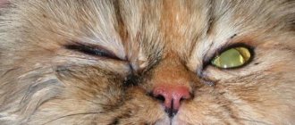
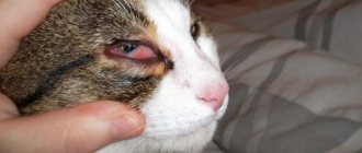
A street cat scratched a dog's eye during a fight.
What should you do in this case? At home, you can prepare an infusion of chamomile or calendula and gently rinse the injured eye with it. To prevent infection of the second healthy eye, it is recommended to rinse it with a fresh swab dipped in chamomile infusion.
If a dirty cat's claw has slightly scratched your eye, your veterinarian will prescribe an antiseptic wash lotion and eye drops to relieve irritation and inflammation. The course of treatment is individual, as it depends on various factors. The animal is under the supervision of a veterinarian for 1-2 weeks. In case of suppuration, experts recommend treating with antibiotic therapy.
Symptoms
Please note that you will not always witness exactly how the incident occurred. At the same time, the scratch may be small and invisible, which will not allow you to immediately examine it. The fact of corneal damage can be detected by focusing on the behavior of the animal. If a cat hits a dog in the eye, the latter will be:
- Whine
- Shake your head frequently
- Rub the area around the eyes with your paw
Of course, it is much better to take your dog to the vet immediately, ideally straight to an ophthalmologist. However, given that not every city has a veterinarian, let alone highly specialized specialists, it is important to know how to provide first aid to an animal yourself.
Getting particles, dust, sand and other small particles from the environment into the eye
Before visiting a veterinarian, it is recommended to rinse the injured eye with saline solution of sodium chloride or warm boiled water. In a veterinary clinic, the doctor treats the affected eye and applies a solution of novocaine (2%) to reduce pain. Then manipulations are carried out to remove the speck from the eye.
To do this, take a swab moistened with a special solution. The veterinarian spreads the eyelids and pours the solution out of the swab into the place where the speck is approximately located. If it is clearly visible, then you can remove the particle with the corner of a cotton swab. After its removal, anti-inflammatory or antimicrobial eye drops are instilled to prevent infection of the damaged organ of vision.