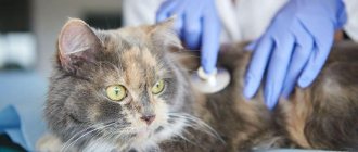
Many cat owners, asking why a cat is breathing with its mouth open, do not suspect that their pet is in serious danger at this moment.
The point is this: the respiratory system of cats is designed in such a way that breathing with an open mouth is uncharacteristic for these animals, unlike dogs. If you see that your animal has begun to breathe heavily, while opening its mouth and hanging out its tongue, this means that it is seriously ill.
If you don't plan to spend a lot of time with your cat, or if you find it difficult to care for an animal with a hereditary predisposition to diseases, you may want to consider other breeds that are less prone to hereditary diseases. You can find them by contacting your local cat lovers club. They will also tell you the rules for caring for a particular breed of animal.
A cat breathes with its mouth open if there is severe oxygen starvation in its body.
Features of breathing in cats
It’s worth starting with the fact that among the reasons why cats switch to this type of breathing, there are physiological ones, that is, those that are normal and pathological ones, caused by various diseases.
One of the cases of physiological mouth breathing in cats on video.
But, of course, this is rather a pleasant and funny case when a cat breathes with its mouth open. But this is not always the case. Mouth breathing, caused by physiological reasons, usually does not last long, immediately after oxygen saturation, the body returns to normal mode. Happens when:
- High ambient temperatures are more common in long-haired and plush breeds.
- And also after intense physical activity, especially in cats with severe obesity.
What to do if pulmonary fibrosis is detected?
Fibrosis is a pathological proliferation of connective tissue, which leads to a decrease in the size of the alveolar vesicles, that is, the air space. With pneumonia, adhesions of connective tissue form around foci of inflammation. As a result, the porous tissue of the respiratory organ becomes denser, the structure of the lung matrix is disrupted, and the space available for air and gas exchange is reduced.
Pulmonary fibrosis resembles scars and requires self-treatment. The consequences of such a complication of pneumonia may be irreversible.
Thus, fibrosis represents an aggressive but physiologically justified response to inflammation or mechanical injury. The body creates an artificial barrier between affected and healthy tissues. Pulmonary fibrosis as an outcome of viral pneumonia caused by COVID-19 is observed in approximately 15% of cases.
As a rule, small local fibrosis in one of the segments of the lung, unlike multiple diffuse fibrosis, does not affect the patient’s quality of life. However, the complication requires separate treatment, which, if necessary, will be prescribed by a pulmonologist after studying the results of CT scans and functional tests.
Manifestations of fibrosis may include shortness of breath, a feeling of lack of air, and intolerance to physical activity. The goal of a rehabilitation course for pulmonary fibrosis is to restore the previous lung volume. The patient is recommended conservative treatment, including drug therapy, breathing exercises, and lifestyle correction.
It is possible to finally assess the severity of fibrotic changes after coronavirus, as well as their significance, only after 12 months. Therefore, it is recommended to repeat a CT scan of the lungs for fibrosis every year.
Causes of mouth breathing
The causes of heavy mouth breathing in cats due to illness are much more extensive. The main function of the respiratory system is to saturate the body with oxygen when inhaling and removing carbon dioxide when exhaling. The upper and lower respiratory tracts, lungs and respiratory muscles participate in this gas exchange.
A disruption in the functioning of any of the components leads to a malfunction in the system and oxygen starvation; in order to compensate for the lack of vital O2, the body has to change the type of breathing. Let us consider in more detail the pathologies that can cause acute respiratory failure. They are divided into two groups: the pulmonary form, which is directly related to diseases of the respiratory system, and the ventilation form, which is a gas exchange disorder caused by extrapulmonary diseases.
| The pulmonary form includes |
|
| Violation of central regulation and patency of the nerve impulse |
|
| Muscle disorders |
|
| Disorders related directly to the chest |
|
| The course of respiratory failure may be |
|
And it depends on the reasons that caused the pathology. One of the most common is pulmonary edema.
“Can’t breathe,” or what you need to know about COPD
Breathing is an amazing process of purifying and absorbing oxygen. And the “shelf life” of the filtering capacity of the respiratory system is designed for decades. However, modern air pollution, smoking and other factors can significantly reduce the time of uninterrupted functioning of the bronchi and lungs. And one of the natural manifestations of a “respiratory breakdown” is chronic shortness of breath and a sharp decrease in quality of life.
When the “filter” fails
COPD, or chronic obstructive pulmonary disease, is also known as chronic bronchitis and emphysema. And for its occurrence, it is customary to “blame” the toxic effects of cigarette smoke and smoke from solid fuels (wood and coal).
Toxic emissions from production, the general dustiness of the air, as well as the “atmosphere” in the room do not go unnoticed, especially when working in hazardous “dust” industries.
However, the influence of harmful factors still does not always lead to the development of irreversible “respiratory” changes, and the formation of COPD is associated with the duration and intensity of the “damaging effect”.
The thing is that the inner lining (epithelium) of the bronchi, which is in direct contact with air, is “superficially” renewed every 2-3 weeks. And complete recovery from serious damage is possible within a year.
It is for this reason that the development of COPD “requires” chronic damage to cells, when the latter do not have time to recover, and the “respiratory filter” becomes powerless. And only in this case does the process of pathological and, over time, irreversible changes begin.
Defense forces work against
Normally, the cells of the bronchi and lungs are reliably “fenced” by a number of protective mechanisms.
First of all, it is a secret (mucus) that mechanically binds foreign objects.
Secondly, coughing as a way to get rid of an irritant using air flow.
And, of course, immune cells that instantly trigger a cascade of protective chemical reactions leading to:
- increased mucus production and increased “stickiness” (phlegm),
- narrowing of the bronchi (spasm), as a way to reduce new “intakes” of irritating substances and, at the same time, increase air pressure during exhalation (cough),
- and increasing the activity of certain enzymes (as a way to “dissolve” the irritant and inflammatory proteins).
Usually these mechanisms are sufficient to cope with the effects of harmful factors and restore the integrity of the mucosa. However, if the influence of irritants is prolonged, the cells do not have time to recover, the area of damage slowly increases, and at the same time the protective mechanisms are decompensated:
- sputum becomes “uncoughable” and accumulates in the bronchi, narrowing their diameter and creating comfortable conditions for the proliferation of pathogenic flora;
- bronchospasm becomes constant, and the “whistle” when inhaling is heard from a distance;
- The elastic tissue of the lungs is destroyed under the influence of inflammatory enzymes, sharply losing the ability to expand and contract, which affects the extensibility of the lung tissue during inspiration.
"Side effects" of COPD
Obstructive pulmonary disease not only significantly worsens the quality of life, but also provokes the development of severe complications, including:
- brain damage (due to chronic oxygen starvation);
- overload of the heart and the development of heart failure;
- and increased risk of developing lung cancer
Among the milder “effects” of COPD are chronic fatigue, deterioration of thought processes, headaches and other consequences of prolonged oxygen deficiency.
At the same time, due to the high ability to recover, the disease develops surprisingly slowly. And, for example, COPD of smokers, as a diagnosis, is established after 30-40 years of smoking. That is, on average, by 50-60 years.
Diagnostics
For early detection of COPD and prevention of severe complications, you should remember that:
- the disease always begins with signs of chronic bronchitis (characteristic morning cough)
- and symptoms similar to anemia (weakness, fatigue, pale skin, decreased immunity, deterioration of the skin, hair and nails, heart rhythm disturbances, and others) against the background of normal, and even increased (as a way of compensation) blood hemoglobin.
The direct diagnosis of “COPD” is only based on the results of an X-ray examination of the lungs (or CT), as well as functional tests (determination of capacity and volume).
And as an additional diagnostic measure, a blood test for alpha-1-antitrypsin can be used.
This enzyme is synthesized by the liver and protects the lungs from elastase.
The latter is produced in excessive quantities by neutrophils (immune cells) during inflammation and serves to “dissolve” certain proteins, which are then freely removed from the lungs.
And with a deficiency of alpha-1-antitrypsin, elastase “goes out of control” and destroys the elastic tissue of the lungs, provoking the development of COPD.
Moreover, in most cases, such a deficiency is of “genetic” origin and manifests itself clinically early, without connection with harmful factors and age.
Therefore, the analysis is primarily indicated for patients under 40 years of age, with complaints of wheezing, chronic cough, shortness of breath after exercise, who do not smoke or are not exposed to dust. And also to calculate the risk of COPD in the future, even if no symptoms are currently observed.
Pulmonary edema in a cat
Pulmonary edema is a condition in which the level of fluid in the lungs is higher than normal. Blood plasma leaves the vessel and fills the intercellular pulmonary space. The lungs increase in size, but can no longer fully perform their direct function.
The reason for oxygen starvation during edema is that with each breath, the accumulated liquid tends to foam. From one milliliter of such liquid, about 15 milliliters of foam are obtained.
As it accumulates, it fills the airways and significantly increases the load on the respiratory muscles. The oxygen that enters the lungs simply does not reach the alveoli, mixing with foam bubbles. As a result, respiratory failure progresses.
Specifics of diseases of purebred cats
In some purebred animals, as well as their mixed breeds, edema can be caused by heart failure. If we talk about breeds, the most predisposed are Scottish Fold, Oriental and Abyssinian cats, Sphynxes, Cornish Rexes and Maine Coons.
Symptoms of pulmonary edema in cats and kittens
Video about the symptoms of pulmonary edema in cats and dogs
As for clinical symptoms and manifestations, at first there is lethargy, complete or partial loss of appetite, and decreased activity of the cat. Over time, anxiety, shortness of breath (the cat breathes through its mouth), and tachycardia appear. Lack of oxygen causes cyanosis of the mucous membranes.
More severe conditions are accompanied by wheezing, initially dry, then wet. White foam appears from the nose and mouth, sometimes mixed with blood. Coordination and heart rhythm are impaired. The outcome of respiratory failure can be a hypoxemic coma, as a result of the death of brain cells due to lack of oxygen and the death of the animal, if it does not occur earlier due to the severity of the condition.
Frequent, difficult breathing through the mouth and cyanosis (blue color of the oral mucosa) require urgent treatment at the hospital for examination by a veterinarian. Since the reason that caused this condition can threaten health and even life.
Do I need to do a CT scan of the lungs again?
Situation 1. You did the first CT scan of the lungs, but too early. Diagnostics showed no signs of viral pneumonia, but after a few days the symptoms of acute respiratory disease intensified - it was difficult to breathe, there was noticeable discomfort in the chest, and an unproductive cough.
A CT scan of the lungs is recommended for every patient infected with coronavirus who has symptoms of acute respiratory disease (shortness of breath, difficulty breathing, cough), high temperature, or decreased blood oxygen saturation. At the same time, you do not need to undergo this examination immediately, but 6-8 days after the onset of symptoms.
If a CT scan is done too early, the first diagnosis may not show pathological changes in the respiratory organ, since the virus has not yet had time to move down the respiratory tract to the lungs and cause damage that provokes swelling, the accumulation of liquid exudate, and the formation of “frosted glasses.” A patient who assumes a relatively safe course of the disease may already feel discomfort and shortness of breath within a few days. The examination will have to be repeated to assess how damaged the lungs are.
If more than 50% of the lungs are affected, hospitalization is required. In this regard, it is important to understand that signs of pneumonia do not become visible immediately.
Situation 2. You are sick, CT shows signs of viral pneumonia (CT-1, CT-2). Positive dynamics are observed during treatment. Complications (fibrous changes, pulmonary edema, honeycomb lung symptom) were not identified.
Repeated computed tomography of the lungs with CT-1 and CT-2 without complications is enough to be done once after a course of treatment, which takes 1-2 months. This will be useful to clarify the results of treatment and the absence of complications. However, if the lung involvement is less than 25% without complications, it is not necessary to repeat the CT scan. A regular x-ray with a small percentage of inflammation is not informative.
Situation 3. You are sick, CT shows signs of viral pneumonia (CT-3, CT-4). Or you have a lower percentage of pneumonia, but fibrous or reticular changes, an interstitial component, or other complications have been identified. Positive dynamics are observed during treatment.
It is recommended to do a control CT scan after a course of treatment for viral pneumonia (after 1-2 months). If necessary, repeat the examination after therapy for pulmonary fibrosis.
Situation 4. You are sick, CT shows signs of viral pneumonia. Treatment does not bring results, health worsens.
The attending physician, familiar with the patient's clinic, may recommend a repeat CT scan of the lungs in 1-2 weeks or earlier, depending on how you feel.
Treatment
When a cat with rapid and heavy breathing enters the clinic, a team of doctors immediately diagnoses the cause of the pathology and takes all necessary measures to stabilize the animal’s condition. The owner must provide all the necessary medical history data, so it is better if the cat is brought to the hospital by a person who can answer all the doctor’s questions. Subsequently, the animal is examined and the necessary diagnostic tests are carried out, including blood tests, x-rays, ultrasound, and ECG.
First of all, intensive therapy is aimed at saturating the body with oxygen. In addition, surgery may be necessary. If there is a large accumulation of fluid in the chest cavity, thoracentesis is performed, which is a puncture of the chest with a thin needle to remove the accumulated fluid. If respiratory failure is caused by injuries, the cause is also promptly eliminated. The prognosis is always different and depends on a timely visit to the clinic, the causes of the disease, the severity of the condition and, of course, the quality of the care provided.
A cat in a car breathes with its mouth open
If you took your pet with you in the car and while driving you noticed that the cat was breathing with its mouth open, most likely the pet was seasick. In principle, this is not so scary, but in the future, before taking the animal anywhere, first prepare for transportation. Key points about transporting a cat:
- do not feed your pet before the trip, but make sure he drinks;
- if the cat in the car begins to breathe with its mouth open, do not disturb the animal, it is better to stop for a while and give the pet a drink;
- do not leave your pet in a car in a parking lot, especially in summer.
Is repeat CT scanning of the lungs harmful?
Any study using ionizing x-ray radiation is a burden on the body. During the year, radiation exposure not exceeding 15-20 mSv is permissible. For health reasons - up to 50 mSv, according to the current Radiation Safety Standards of the Russian Federation (NRB-99). When treating a disease with a high mortality rate and complications, such as viral pneumonia caused by COVID-19, repeat computed tomography is more beneficial than harmful. Expedient radiation exposure is justified.
Respiratory diseases in cats
The group of respiratory diseases of cats includes infectious diseases such as:
- rhinotracheitis;
- calcivirosis (calicivirus infection);
- mycoplasmosis;
- chlamydia.
Making a diagnosis is complicated by the fact that these diseases occur with similar symptoms, and in addition, often together. Thus, one cat can develop several infections at once.
Caring owners usually immediately notice that their cat is breathing with her mouth open and sneezing, she has nasal discharge and lacrimation, or wheezing and wheezing can be heard from a breathing pet. In this case, the general condition of the animal remains the same or a slight lethargy appears. If your cat has completely lost its appetite, you should immediately contact your veterinarian.