Eye diseases in cats deserve special attention, as they often cause deterioration or complete loss of vision. Despite the development of other sense organs, blindness significantly worsens the animal’s quality of life. You can avoid troubles with the help of prevention and timely treatment, started immediately after the detection of alarming symptoms.
How can you tell if something is wrong with your cat's eye?
If there is something wrong with a cat's eye, it is easy to understand by its condition. The following symptoms will indicate the disease
:
- increased lacrimation and discharge of pus;
- redness of the mucous membrane and inflammation of the eyelids;
- photophobia, covering of the third eyelid and frequent blinking;
- pain on palpation;
- the appearance of itching;
- sticking of the eyelids after waking up due to the drying out of the accumulated exudate.
Depending on the cause of the malaise, symptoms may be supplemented by worsening appetite, apathy, severe thirst and fever. If you notice at least a few of the listed signs, be sure to schedule your pet for an examination at a veterinary clinic.
How to live with a cat if allergies are confirmed?
Based on the diagnostic results, it turned out that your body reacts specifically to the cat, but you do not want to part with your pet. What to do? By following a number of recommendations, you can reduce the likelihood of exacerbation of allergies:
- Try to pet the animal less, do not kiss it. After being held, wash it with soap.
- Regularly give your cat water treatments and brush her
- Replace the open tray with a closed one
- Do wet cleaning as often as possible, at least twice a week
- Don't forget to ventilate the rooms several times a day.
- Air washers will help clean the room from flying microparticles
- If possible, remove all carpets, bedding, and soft toys. Put things in the closet immediately
These rules will help you get along with your tailed friend in the same house. But their observance requires care and patience.
Causes of eye problems in cats
Three groups of reasons are to blame for the occurrence of ophthalmic diseases. These include:
- infectious, due to the negative effects of viruses and bacteria;
- non-infectious, including tumors, injuries and allergies;
- congenital, associated with breed predisposition and poor genetics.
The risk group includes extreme-type Persians with pronounced breed traits. Due to increased tear production, which forms pathogenic microflora, their tear ducts are very often infected with bacteria.
Types of eyelid diseases and their treatment
Symptoms and methods of treating eye diseases in cats depend not only on the cause, but also on the area of the lesion. The main blow is always taken by the eyelids, which protect the more delicate layers from the aggressive influence of the external environment.
Blepharitis
Staphylococcal infections that enter the eye cause unilateral or internal blepharitis in cats. This disease is accompanied by severe redness and inflammation of the corners of the eyelid, as well as the discharge of purulent exudate. Dried pus gradually covers the entire affected area, causing thickening and ulceration of the skin.
In addition to infection, blepharitis can be caused by allergies or demodicosis. Inflammation is treated with antibiotics, antihistamines or acaricidal drugs.
Third eyelid prolapse
This term refers to the transparent nictitating membrane, which performs a protective and moisturizing function. It is located in the inner corner of the eye next to the nose. Its loss is accompanied by uncontrollable twitching of the eyelids, lacrimation and copious discharge of pus or mucus.
Pathology develops with infections, allergies, parasitosis, foreign objects and fusion of the eyelids with each other. To reduce it, they resort to surgery.
Entropion of the eyelids
An alternative name for this disease is entropion. Most often it affects the lower eyelid, causing photophobia and excessive lacrimation. With entropion, it is very easy to understand that something is wrong with the cat’s eye, because she experiences severe pain due to prickly eyelashes and hairs.
The pathology is caused by congenital disorders, injuries and neoplasms. The problem is eliminated by excision of excess skin and formation of the correct palpebral fissure.
Ptosis
Characterized by involuntary drooping of the upper eyelid. The diseased eye opens only partially, and the palpebral fissure narrows. Ptosis occurs as a result of complications of inflammatory processes, paralysis of the facial nerve or weakness of the orbicularis muscle covering the anterior parts of the orbit. It is treated exclusively by surgery.
Lagophthalmos
External signs of lagophthalmos are similar to ptosis. The difference is that the eyelids cannot close. Because of this, the cat has to sleep with its eyes open. The causes and treatment are the same as ptosis.
Fusion of the eyelids with each other or with the conjunctiva
The first type of disorder is called ankyloblepharon, and the second - symblepharon. These pathologies develop from birth, with chronic blepharitis and as a result of mechanical damage. Both cases are accompanied by tissue scarring and the inability to open the eyes. To eliminate the fusion, the help of a surgeon is required.
Blepharitis
Inflammation of the eyelids, which is quite common in furry cats, is divided into three types:
- Scaly blepharitis (also called simple);
- Ulcerative blepharitis;
- Meibomian blepharitis.
Symptoms differ for each type, but there are common ones that are always noticeable. Namely:
- Itching;
- Edema;
- Redness;
- Tearing.
With scaly blepharitis, you can notice a clear thickening of the eyelids, after which gray scales appear there. Over time, the eyelashes will begin to fall out, and pus will be released in place of the scales.
If treatment is not started, scaly blepharitis can quickly turn into ulcerative blepharitis. With it, crusts of pus dry out, fall off, and in their place sores open. The inflammation gets worse. Scarring may cause the skin to tighten.
Meibomian blepharitis is similar to a stye. The eyelid swells and turns red. Pus begins to ooze out profusely.
Pathologies of the fundus and lens
Ophthalmic diseases in cats can affect the fundus and lens. These organs are responsible for visual acuity, so when they are damaged, it becomes more difficult for the animal to catch objects at a distance.
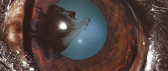
Cataract
It is characterized by a loss of transparency of the lens, impairing the ability to refract light rays. The main symptoms of cataracts are clouding of the pupil and iris, as well as a gradual change in their shade to bluish-gray.
The causes of the disease lie in congenital disorders, advanced infections, injuries and diabetes. At the initial stage, only damaged particles are removed, and at an advanced stage, the entire lens is removed at once.
Glaucoma
Accompanied by the appearance of a greenish cataract, redness of the mucous membrane and an enlarged pupil. The main cause of glaucoma is increased intraocular pressure. The pathology cannot be treated, but thanks to cryotherapy and antiglaucomatous drugs, its development can be slowed down.
Bleeding in the eye
Atherosclerosis
Diabetes
Fungus
26717 March 22
IMPORTANT!
The information in this section cannot be used for self-diagnosis and self-treatment.
In case of pain or other exacerbation of the disease, diagnostic tests should be prescribed only by the attending physician. To make a diagnosis and properly prescribe treatment, you should contact your doctor. Hemorrhage in the eye: causes of occurrence, what diseases it occurs with, diagnosis and treatment methods.
Definition
Hemorrhage in the eye is the accumulation of blood under the conjunctiva, in and between the membranes of the eye, in the chambers of the eye or in the vitreous body. Hemorrhage is caused by a violation of the integrity of the blood vessels of the eye.
The eyes continuously perceive, transform and send a huge amount of information to the visual analyzer.
The eyes are sensitive to changes in the body and, although quite protected, are easily damaged by external influences.
The eyeball is located in the cavity of the orbit - a bone frame; the orbit protects the eyeball from above, below, behind and on the sides. The eye in the orbit is located in fatty tissue - a kind of pillow. In front, the eyeball is protected by eyelids and eyelashes.
Eyeball
consists of three shells:
- external, represented by a convex transparent cornea, turning into the sclera - the frame of the eyeball;
- middle - in front is the colored iris, then the ciliary body, and in the back is the choroid itself, represented by a network of small arteries and veins. At the center of the iris is the pupil, which allows light to enter the eye. The ciliary body produces intraocular fluid, which is needed to maintain the shape of the eye and help with metabolism. The lens is attached to the ciliary body with the help of muscles and ligaments. By contracting and relaxing the muscles, the curvature of the lens changes, thus the eye “switches” the distance and focuses the image;
- the inner retina, which contains rods and cones that perceive light and convert it into an electrical impulse, which is then sent to the brain.
Vitreous body
fills the space between the lens and the retina and is a gel-like substance.
The eye has two spaces filled with intraocular fluid: the anterior chamber of the eye (between the cornea and the iris) and the posterior chamber of the eye (between the iris and the vitreous body, which contains the lens).
The cameras communicate with each other through the pupil. Conjunctiva
- This is the mucous membrane, it lines the inside of the eyelids and extends to the visible part of the eye. The conjunctiva is nourished by a network of very small vessels located directly underneath it. Externally, it is constantly washed with tears produced by the lacrimal glands.
Types of hemorrhages in the eye
Hemorrhages in the eye are divided depending on the place where the blood accumulates:
- With hyposphagma, blood accumulates between the conjunctiva and sclera due to damage to the conjunctival vessels. It looks like a bright red spot on the white of the eye. This is the least dangerous type of hemorrhage and does not lead to visual impairment, but you should be wary if it occurs regularly;
- With hyphema, blood flows into the anterior chamber of the eye. Hyphema has 4 degrees: 1st degree is characterized by filling the chamber by no more than 1/3, 2nd – by no more than 1/2, 3rd – by more than 1/2, but not completely, 4th – complete filling of the anterior chamber of the eye with blood. If blood reaches the pupil, the vision becomes blurred, vision decreases, and fear of bright light appears. With this type of hemorrhage, pain occurs;
- with hemophthalmia, blood fills the vitreous body. There are complete and incomplete hemophthalmos. Vision drops sharply, the degree of impairment of which depends on the volume of blood shed. A very dangerous type of hemorrhage that can lead to filling of the vitreous with connective tissue, permanent vision loss and possible retinal detachment;
- In retinal hemorrhages, blood accumulates just in front of the retina (preretinal hemorrhages), inside the retina (intraretinal hemorrhages), or just after the rod and cone layer (subretinal hemorrhages).
This is the most serious type of hemorrhage, most often leading to decreased or even loss of vision.
Possible causes of hemorrhage in the eye
The most likely causes of hemorrhage in the eye include:
- mechanical impacts, eye injuries, head injuries - the degree of damage depends on the force of the impact;
- intraocular operations;
- rupture of newly formed vessels;
- diseases and conditions that affect the composition and properties of blood, disrupting the blood clotting process (taking blood-thinning drugs, hematological diseases);
- diseases affecting the vascular wall (hypertension, atherosclerosis, diabetes mellitus);
- eye diseases: glaucoma (with increased intraocular pressure), high degree of myopia, retinal detachment, thrombosis (blockage) of the central retinal vein;
- various infections (viral, bacterial, fungal, etc.) leading to inflammation of the eye structures. Conjunctivitis (inflammation of the conjunctiva), iritis (irises), iridocyclitis (iris and ciliary body), chorioretinitis (choroid and retina), retinitis (retina only);
- increased intracranial pressure due to excess volume in the cranial cavity due to intracranial hematomas, brain tumors, cerebral edema;
- an increase in intra-abdominal and, as a consequence, venous pressure during severe physical exertion, debilitating vomiting, and during childbirth.
For non-penetrating blunt eye injuries
The cause of vascular rupture is a sharp increase in intraocular pressure, the same mechanism is triggered in glaucoma.
Newly formed vessels have a rather fragile wall; they can appear without good reason or to replace damaged ones, for example, in diabetes mellitus
. In addition, an increase in blood sugar levels leads to damage to the smallest vessels - capillaries.
For infectious diseases
the inflammatory process leads to increased vascular permeability, which can cause damage.
Blood diseases that most often lead to hemorrhages include hemorrhagic diathesis
.
This is a group of diseases in which the processes of stopping bleeding are disrupted.
For example, in hemophilia, there is a hereditary decrease in blood clotting factors, which prevents a blood clot from forming. In thrombocytopenia, there are not enough platelets to initially block the lesion. Oncological diseases of the blood. The most striking example is acute leukemia
. Tumor cells displace bone marrow cells, then enter the bloodstream and replace all healthy cells, including those responsible for stopping bleeding.
Atherosclerosis
manifested by the deposition of cholesterol plaques in blood vessels.
A constant or periodic increase in blood pressure
damages the inner layer of blood vessels - the endothelium, while the middle, muscular layer thickens, and the vessel loses its elasticity. These changes sooner or later lead to a violation of the integrity of any vessel.
Retinal disinsertion
from the choroid with hemorrhage occurs with eye injuries, inflammatory diseases of the retina, and disturbances in its nutrition. The risk of detachment increases in people with a high degree of myopia.
Symptoms of retinal detachment include a sharp decrease in vision, narrowing of the visual field or loss of a portion of the image, the presence of spots in front of the eyes, flashes of light.
Which doctors should you contact if there is a hemorrhage in the eye?
The treatment of hemorrhages in the eye is carried out by an ophthalmologist; to correct concomitant conditions, a consultation with a general practitioner may be required.
Diagnosis and examinations for hemorrhage in the eye
The doctor conducts a survey and examination of the patient and his eyes, determines visual acuity, measures blood pressure, intraocular pressure, studies the structures of the anterior chamber of the eye, and examines the fundus, if possible.
A number of additional examination methods may be required:
- ultrasound examination of the eye;
- electroretinography to assess the functional activity of the retina;
- general blood analysis;
Diseases of the cornea and mucosa, appropriate treatment
The cornea (cornea) acts as a refractive lens, the mucous membrane (conjunctiva) protects the visual organ from drying out, and the vascular membrane (uveal tract) saturates the retina with useful microelements. All these organs are very vulnerable, so after damage to the eyelids, they are the ones who suffer from the effects of negative factors.
Conjunctivitis
If a cat's eye is inflamed and swollen, and the conjunctiva has acquired a bright crimson hue, then most likely he has conjunctivitis. Its appearance is associated with infections and allergies, so antibiotics or antihistamines are used for treatment. If there is an abundant accumulation of pus and the presence of multiple follicles, surgery is performed.
Uveitis
With uveitis, a cat's eyes hurt a lot, so she constantly closes her eyelids and avoids bright light. Advanced inflammation of the uveal tract rarely responds to drug treatment, so the only salvation is removal of the affected organ. In the early stages, it is enough to eliminate the root cause and undergo a course of symptomatic therapy.
Keratitis
The main cause of inflammation of the cornea in cats is similar to previous pathologies. Most often, keratitis develops when infected with bacteria and viruses, so the victim is prescribed antibiotics or antivirals. In advanced cases, laser correction is used.
The inflamed cornea becomes blue or gray and dull, and the mucous membranes become noticeably red. A sick pet tries to avoid bright light and constantly rubs its face with its paws. Without timely help, keratitis can be complicated by cataracts or glaucoma.
Keratitis
Keratitis is a lesion of the cornea of the eye. It can be caused by either trauma, including foreign body impact, or infection. The main symptoms of keratitis are:
- photophobia;
- profuse lacrimation;
- corneal clouding;
- accumulation of discharge in the corner of the eye.
When treating keratitis, it is important to exclude the possibility of its relapse : eliminate the cause of the disease. Effective drug intervention is impossible without the help of a veterinarian. Only a specialist can correctly determine the cause of the disease and prescribe the correct therapy.
Other pathologies
Less common eye diseases in cats include the following:
:
- Canaliculitis
, or inflammation of the tear ducts. Occurs when the ducts become clogged and is accompanied by profuse lacrimation. It is treated by washing the clogged lacrimal sac or removing it.
- Exophthalmos (proptosis) and enophthalmos
, or prolapse and retraction of the eyeball. Changes are visible to the naked eye. The organ is returned to its original position under general anesthesia.
- Panophthalmitis
. A most dangerous pathology that affects all eye tissues. Accompanied by an enlargement of the eye and the discharge of pus. To avoid brain infection, the affected organ is removed.
- Adenoma of the third century
. It is characterized by severe redness and thickening of the third eyelid as a result of inflammation of the lymph nodes. This makes it look like a cherry. The organ affected by the adenoma is sutured to its normal state or removed.
Due to the high likelihood of complications, it is better to entrust treatment to a veterinarian. Self-administration of drugs rarely brings the desired result. At best, you will simply smooth out the existing symptoms and complicate the diagnosis.
Diagnosis of the disease
If your pet constantly closes its eye, a veterinarian will find out the reasons. If alarming symptoms are detected, it is necessary to take the cat to the clinic.
Diagnostics includes procedures such as:
- visual acuity test;
- assessment of the general condition of the body;
- examination of the visual organs for damage;
- comparison of symmetrical features (size and shape of pupils, eyelids, eyeballs and slits);
- Ultrasound of the affected area;
- staining test;
- CT or MRI of the brain;
- X-ray of the skull.
Ophthalmological pathology may be a consequence of traumatic brain injury. Therefore, the examination must be comprehensive.
It is recommended to donate blood, urine and feces for analysis. Problems with the eyes can be caused by pathologies of internal organs or parasitic diseases.
When veterinary help is urgently needed
If something is wrong with a cat’s eye, the owner can provide first aid. After this, it is recommended to consult a doctor. This must be done even if the problem seems completely insignificant. After all, it is impossible to make a diagnosis on your own, and your mistake can result in premature blindness of your beloved pet.
Emergency assistance will be required if pus appears, high fever, signs of cataracts and glaucoma, burns or other health-threatening injuries. In these cases, it is recommended to contact the nearest 24-hour veterinary clinic without waiting until morning.
First aid and prevention
Why do my cats always have red eyes? The reason is different in each specific case. The syndrome may be a consequence of violation of the rules for caring for the animal, or it may be a symptom of a serious illness. Therefore, if the redness does not go away for a long time, you should not delay a visit to the veterinarian. Only he can make a high-quality diagnosis, assess the pet’s condition and determine further treatment tactics.
If the owner does not have the opportunity to immediately go to the clinic, you can use first aid techniques for ophthalmological problems. They will help alleviate the pet’s condition somewhat and, in some cases, have a therapeutic effect.
- It is necessary to put a protective collar on the neck of a cat or kitten, which will prevent them from touching their eyes with their paws, and therefore prevent the risk of secondary infection.
- You should regularly remove discharge from the eyes and rinse them with cotton pads soaked in warm boiled water or a strong solution of tea leaves.
- To reduce the inflammatory reaction, you can instill antiseptic eye drops or an “artificial tear” drug.
- Hair hanging over the eyes should be trimmed more often so that it does not injure the cornea.
If you ignore a symptom such as redness of the eye sclera, you may miss the onset of a serious illness in your cat. It’s better to play it safe and take your pet to the veterinarian as soon as possible for an examination and consultation, so that you don’t have to regret the lost time later.
Treatment at home
After the diagnosis is made, the mustachioed patient must be provided with the most comfortable conditions possible. Place it in a darkened room, away from noise sources, other pets, children and other irritants.
To avoid further injuries, be sure to trim your cat’s claws and do not let him outside until he recovers. In addition to the basic treatment, which includes the use of eye drops and ointments, you will need to pay close attention to hygiene through rinsing.
Washing and hygiene
Washing is carried out using cotton pads, guided by the following algorithm
:
- Prepare all the necessary tools and preparations in advance to reduce the risk of traumatizing the animal with painful waiting.
- Wrap your pet in a thick cloth or find an assistant who can hold him during the procedure.
- Soak a cotton pad in the medicinal solution, squeeze it out and open the cat's eyelids.
- Gently move the disc over the eye, moving from the outer corner to the inner.
- Repeat the previous steps with the other eye, not forgetting to change the cotton pad.
The procedure can also be carried out using a syringe without a needle. To do this, you need to irrigate each eye with the solution under low pressure and gently blot them with a clean, lint-free cloth. After removing the accumulated dirt and soaking the crusts, you can begin applying the main preparation.
Drops and ointments
If something is wrong with a cat’s eye, then special drops and ointments will be required to normalize its condition. The list of necessary medications will depend on the cause of the disease:
- bacterial infection – Iris, Leopard, Levomycetin;
- viral infection – Anandin;
- fungal infection – Tetracycline;
- injuries - Traumeel.
The drops are instilled into both eyes at once, and the ointment is applied only to the disturbing area. After application, close the eyelids tightly and gently massage so that the product is evenly distributed inside. During treatment, it is recommended to wear an Elizabethan collar on your pet so that it does not inflict further injuries on itself when scratching.
Is it possible to use folk remedies?
Folk remedies can only be used as part of complex therapy. They do not fight the root cause, but only alleviate the patient’s condition by eliminating associated symptoms. It is also important to note that some herbs can cause an allergic reaction, so it is best to use them only under the supervision of a veterinarian.
Please note that the popular recipe with olive oil is extremely dangerous. Any oily substances create a very dense film. Because of this, new bacteria can quickly grow on the mucous membrane after treatment.
Features of treatment of pregnant, lactating and young individuals
Kittens, as well as pregnant and lactating animals, need gentler medications and lower dosages. Their immunity is weakened, so most antibiotics cause severe complications. Some of them can lead to miscarriages or stillbirths.
They also resort to surgery with extreme caution, since the anesthesia used can affect the course of pregnancy or the baby’s fragile heart. The indication for its implementation is a serious threat to life that exceeds the existing risks.
Eye drops and ointments for allergic conjunctivitis
In case of allergic conjunctivitis, it is necessary, first of all, to eliminate or minimize contact with the allergen. To relieve symptoms, antihistamine H1-blockers are prescribed to suppress the allergic reaction:
- drops Allergodil, Opatanol, Cromohexal, Lecrolin, Ketotifen, etc.;
- decongestant and vasoconstrictor drops (Vizin, Visomitin, Oksial);
- eye ointments, incl. hormonal (Hydrocortisone, Dexamethasone);
Tablets of systemic antihistamines (for example, Tavegil, Suprastin, Loratadine and their analogues) may also be prescribed.