A hernia in a cat is a protrusion of internal organs or parts thereof from the cavity that was normal for them. The hole can be natural or formed as a result of pathology, ruptures of the serous membrane or muscle tissue. The bulging occurs under the skin, into internal cavities, the space between muscles or skin pockets.
It can threaten the life and health of a pet, since when internal organs are infringed, it often leads to necrosis and sepsis. There are several types of abdominal hernias in cats, and are divided into umbilical, inguinal and intermediate. The pathology has no breed, gender or age restrictions, and can be diagnosed in both kittens and adults.
What is a hernia in cats and kittens?
This term refers to a disease that involves the loss of internal organs or parts thereof through natural or pathologically formed openings. In addition to neighboring cavities, they can fall out into the subcutaneous or intermuscular space.
Education itself always consists of 3 parts:
- hernial contents - an organ or part of it protruding through adjacent tissues;
- hernial sac - stretched tissue of the peritoneum or other cavity where the organ should be located;
- hernial orifices are openings through which an organ protrudes.
Lack of timely treatment is fraught with inflammation or strangulation of the protruding fragment. In both cases, it may suffer from necrosis and lose its functionality.
Surgical reduction of traumatic hernias
When purchasing a puppy, all veterinarians recommend paying attention to the presence or absence of a hernia. So what are hernias, what are they, where do they come from, what to do with them? These are the questions that potential owners and breeders ask themselves if they are faced with hernias in one way or another.
Hernia
is a temporary or persistent prolapse of internal organs through a natural or pathological opening with protrusion of the membrane lining the anatomical cavity. A hernia has its own structure: a hernial ring (opening), through which a hernial sac protrudes, consisting of the peritoneum, transverse abdominal fascia and hernial contents (intestines, omentum, stomach or other organs, tissues).
What types of hernias are there?
Based on their origin, hernias can be congenital or acquired. Congenital
hernias
develop due to anatomical openings that are too wide, or due to a defect in the abdominal wall (Fig. 1).
Figure 1 – Congenital hernia in a puppy.
Acquired hernias
are most often formed due to excessive physical tension of the muscles (Fig. 2).
Figure 2 – Acquired abdominal hernia in a horse.
A hernia in which the contents are freely reduced is called reducible.
, if reduction is impossible due to adhesions, it is called
irreducible
, in the presence of an acute inflammatory reaction -
strangulated
.
Sudden strangulation of a hernia can occur after sudden movements and jumps due to a sharp increase in pressure in the abdominal cavity. Some part of the intestine gets into the hernial sac, and as a result the loop becomes pinched from the outside. According to the location of the hernia, there are the following: umbilical, inguinal, perineal, traumatic, abdominal, diaphragmatic, etc.
UMBILICAL HERNIA.
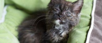
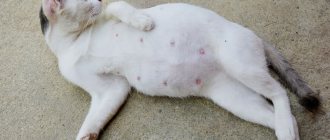
If an umbilical hernia is present in a kitten or puppy, a small, soft to the touch, and painless swelling in the navel area is most often detected. Upon palpation, the contents can be easily reduced into the peritoneum and the hernial ring is clearly palpated. The contents of the hernial sac in an umbilical hernia are most often the omentum, sometimes intestinal loops. The omentum most often does not reduce, as it firmly fuses with the hernial sac. The size of the umbilical hernia can vary from several millimeters to several centimeters (Fig. 3).
Figure 3 - Umbilical hernia in a kitten.
This type of hernia occurs quite often, due to insufficient closure of the abdominal wall near the navel. Most often, an umbilical hernia is a congenital pathology and is a defect in the intrauterine formation of the umbilical ring. Less commonly, in the presence of some anatomical predisposition of the animal, an umbilical hernia occurs due to excessive stress, tension in the abdominal wall and increased intra-abdominal pressure, for example, due to excessive flatulence, severe and constant overeating, and other factors.
Why are umbilical hernias dangerous?
As the animal grows, the hernial sac can increase in volume, while remaining narrowed in the area of the hernial orifice. In most cases, the hernial contents are the omentum, which, however, is sometimes reduced, and sometimes not reduced, due to the fact that it is attached to the hernial sac. Thus, the so-called strangulated hernia can occur, when intestinal loops filled with gas or contents fall into the hernial sac and are strangulated by the hernial ring, intestinal motility is disrupted, obstruction of the gastrointestinal tract develops, which leads to inflammation, further to poor circulation and necrosis (death). Strangulated hernias without surgical intervention are fatal.
ATTENTION!!!
Do not try to reduce a strangulated hernia yourself and under no circumstances use a heating pad!
Hernias do not resolve on their own; gluing nickels and the like to the hernia site does not lead to the desired results!
What kind of help do animals with umbilical hernia need?
If an umbilical hernia is strangulated, then immediate surgery is required, but is it worth bringing your pet to this state?! If the hernia is small, no more than a few millimeters, it may well close on its own without surgical intervention. Those puppies and kittens that have a hernia at birth need to be constantly monitored, i.e. feed fractionally, watch how the hernia behaves (increases in volume or not), visit a veterinarian, since as the pet grows, the formation of the abdominal muscles of the hernial ring also occurs.
Reduction of a non-strangulated umbilical hernia is a planned operation. It is performed under general anesthesia or local anesthesia. Don't wait until the hernia is strangulated, have surgery right now.
The operation is performed in the dorsal position; local anesthetic is injected around the base of the hernia into the abdominal wall from four points (Fig. 4). In this case, the solution is first injected into the subcutaneous tissue, then the needle is penetrated into the dorsal wall of the rectus abdominis vagina, and, piercing it, the needle is immersed in the retroperitoneal tissue, continuing to inject the solution. If the size of the umbilical ring allows you to insert a finger into it, then with the tip of the latter, placed under the edge of the ring, you can control the depth of insertion of the needle, preventing it from penetrating into the abdominal cavity.
Figure 4 - Infiltration of tissues surrounding the hernia.
Having completed anesthesia, the skin is cut with a longitudinal incision in the middle of the hernia. Then, using a gauze swab, a piece of peritoneum is dissected from the skin to the umbilical ring and inserted into the abdominal cavity. A loop-shaped suture made of absorbable material is placed on the hernial ring. It is recommended to scarify the edge of the hernial ring in order to cause a more dense fusion in the future.
For an irreducible hernia, the serous sac is opened. If, upon examination of the hernial contents, a commissure is found between the intestine and the peritoneum, it is necessary to excise the adherent area of the serous sac, leaving it on the intestine. When there is adhesions connecting all the loops of intestines that were in the hernia cavity into a common mass, it is necessary to expand the umbilical ring with a button scalpel, and then push the intestines into the abdominal cavity. The umbilical ring is closed, as in the treatment of a reducible hernia.
In case of a strangulated hernia, as in the previous case, the serous sac is dissected, the strangulated intestine is removed onto a sterile napkin and its condition is determined. In case of swelling and hyperemia, the intestine must be moved into the abdominal cavity. If it is impossible to straighten it with little effort, it is necessary to expand the umbilical ring under finger control with a button scalpel. After repositioning the intestine, the serous sac is amputated. With severe swelling of the intestinal wall, its flabbiness and in some places necrosis beginning, the affected area is resected.
In some cases, when the hernial ring is very wide and the aponeurosis of the abdominal muscles is weak and there is a risk of a hernia forming again, plastic surgery using a polypropylene mesh is used. The polypropylene mesh grows into the tissues of the body and animal and remains in the body for life.
All of the above operations for umbilical hernias end with the removal of excess skin and closure of the skin wound with an interrupted suture.
INGUINAL HERNIA.
The hernial opening in an inguinal hernia is an unnaturally wide gap (the inguinal canal) or a rupture in the tissue of the abdominal wall. Any hernial opening in animals tends to expand over time. The most important anatomical element in an inguinal hernia in animals, which determines the formation of an inguinal hernia, is the natural connection of the cavity of the common vaginal membrane with the abdominal cavity. It communicates through the vaginal canal, which remains after the testis descends into the scrotum. The hernial sac is formed by the parietal peritoneum with the underlying fascia. With prolonged existence, the sac grows with scar tissue, contributing to the formation of adhesions, bridges and the formation of communicating chambers. Inguinal hernias can be congenital or acquired. Acquired hernias are formed due to mechanical damage to the abdominal wall and prolonged straining. An inguinal hernia in a kitten can form due to increased intra-abdominal pressure, when weak spots in the abdominal wall are stretched (the inguinal canal). According to the condition of the contents, there are: reducible, irreducible and strangulated inguinal hernia. Most often, an inguinal hernia is left-sided.
A hernia in which the intestinal loop or omentum (bladder) is located in the vaginal canal should be called a vaginal canal hernia. If the same organs lie in the cavity of the common tunica vaginalis, such a hernia should be called intravaginal. If a new hernial sac is formed, lying completely separately in the scrotum - between the skin, Cooper's fascia, on the one hand, and the common vaginal membrane, on the other, then the hernia should be called true scrotal. In cases where a rupture of the wall of the inguinal canal occurs and the peritoneal hernial sac with its contents lies outside the vaginal canal, i.e., in the inguinal canal, a true inguinal hernia is formed (Fig. 5). This hernia can easily become a true scrotal hernia if the hernial sac descends under the influence of the contents and intra-abdominal pressure to the base of the scrotum.
Figure 5 - Inguinal hernia in a male dog.
An inguinal hernia in females may contain: intestines, bladder or uterus. The first signs of an inguinal hernia are the presence of a small swelling in the groin area on one or both sides, spherical or elongated (Fig. 6.). If the contents of the hernia are a pregnant uterus, then as it grows, the hernial sac increases (in this case, only surgical treatment is meant, with the removal of the fetus).
Figure 6 - Inguinal hernia in a bitch.
ATTENTION!!!
All types of inguinal hernias require surgical intervention. The intervention technique may vary, depending on the sex of the animal and the owner’s wishes to remove or preserve the pet’s testicle. As a rule, in male dogs, the operation is performed to remove the testis. If the testis is left, there is a risk of re-formation of a hernia or necrosis of the testis, that is, another surgical intervention.
ABDOMINAL HERNIA.
The causes of the formation of such hernias are injuries to the abdominal wall with the formation of a muscular or aponeurotic defect. The contents of the hernia, depending on its location, can be any movable organ of the abdominal cavity. Hernias are located in the area of the ilium, xiphoid cartilage, navel, hypochondrium.
Why can it be difficult to recognize an abdominal hernia?
Recognizing a hernia is initially difficult due to the presence of bruise symptoms (diffuse inflammatory edema and hemato-lymphatic extravasation). After the disappearance of inflammatory phenomena (after 8-10 days), ruptures and painless, soft, limited swelling are visible. By pressing on the hernia, its reducibility is established, after which the size of the hernial opening is determined by palpation. The size of the hernia can vary significantly.
The prognosis for a small hernia in the upper part of the iliac region is favorable, since such a hernia does not have a painful effect on the body and is only a beauty defect. If the hernia is located in the lower abdomen and if there is a large hernial opening, the prognosis is questionable. Surgical treatment consists of incising the skin, separating and repositioning the serous sac into the abdominal cavity, and placing a loop-shaped or knotted suture on the edges of the hernial ring. The edges of the hernial opening are scarified, and in some cases a polypropylene mesh is applied (Fig. 7), a procedure called retromuscular repair.
Figure 7 — Application of a polypropylene mesh on the surface of the hernial ring (1-polypropylene mesh, 2-muscles).
PERINEAL HERNIA.
This type of hernia occurs quite often in dogs. It is formed in males due to stretching of the recto-vesical sac of the peritoneum, and in females - the recto-vaginal sac. The contents of a hernia can be: intestines, uterus or bladder. Such hernias are formed due to increased intra-abdominal pressure. Intra-abdominal pressure increases with constipation, vomiting, and straining.
How does the disease manifest?
The disease in males manifests itself as a single (central) or bilateral elastic swelling located under the anus (Fig. 8), and in females - under the genital opening. If the animal is lifted by the thoracic limbs, the volume of the hernia increases, and if by the pelvic limbs, it decreases or disappears completely. Almost no infringement is observed.
Figure 8 - Perineal hernia in a dog.
What is the treatment?
For small hernias, treatment is not required. For hernias of significant size, surgical intervention is necessary, which consists of dissecting, twisting, ligating and suturing the peritoneal hernial sac, freed from the contents, to the posterior edge of the sacroisciatic ligament. If there is a bilateral hernia, both hernial sacs are ligated and sutured to the ligament. In some cases, colorectopexy is additionally performed. The skin wound is closed with a knotted suture, and to protect against contamination with fecal matter, it is lubricated with some kind of disinfectant ointment.
ATTENTION!!!
A complex intervention is performed only if there are no contraindications under anesthesia. A preliminary diagnosis of cardiac pathology is carried out and a blood test (general and biochemical) is taken.
FEMORAL HERNIA
occurs due to the entry of the intestinal loop or omentum, together with the parietal layer of the peritoneum, under the inner fascia of the thigh - into the femoral canal. The disease is manifested by the presence of a soft, painless swelling on the inner surface of the thigh (in the area of the femoral canal). The animal puts its leg on the side on which the hernia is located, and sometimes a splayed gait is noted.
What is the treatment for a femoral hernia?
Treatment
comes down to surgical intervention. It consists of a longitudinal dissection of the skin and underlying parts, preparation of the hernial sac and ligation after repositioning the contents of the hernia into the abdominal cavity, suturing the inner fascia of the thigh and skin. When performing an operation, it is necessary to remember that the femoral vein and artery, and the femoral nerve pass through the femoral canal.
DIAPHRAGMAL HERNIA
in animals, this is a displacement of the abdominal organs into the thoracic cavity, through acquired or congenital damage to the diaphragm. Since the pressure in the abdominal cavity is stronger, the diaphragm bulges towards the chest cavity, therefore the organs are displaced into the chest cavity, and not vice versa (Fig. 9). In addition to separating the cavities, the diaphragm is a support for adjacent organs, and also performs a dynamic function: respiratory (participation in breathing), cardio-vascular, motor-digestive, lymph circulation. In addition, it is the diaphragm that is responsible for inhalation; at rest it provides up to 90% of the tidal volume. Diaphragmatic hernias are divided into traumatic and congenital.
Figure 9 - Diaphragmatic hernia: 1 - small intestine; 2 - liver; a - heart; 4—diaphragm; 5 - gallbladder; c - stomach.
Congenital diaphragmatic hernia
can be pleuroperitoneal or pericardio-pleuroperitoneal. In general, congenital pleuroperitoneal hernias are rare, and puppies with large diaphragmatic defects usually die at birth or shortly thereafter. Congenital pericardio-pleuroperitoneal diaphragmatic hernia is a more common disorder, to which Weimaraner dogs and Persian cats are most susceptible.
With a sliding hernia
due to weakening of the esophageal-diaphragmatic ligament, part of the esophagus and stomach are displaced upward - into the mediastinum. In this case, the fold of the peritoneum forms a hernial sac. The main complication of such a hernia is the straightening of the angle between the esophagus and the stomach, which causes the development of reflux esophagitis (reflux - reflux; esophagitis - inflammation of the esophagus). Sliding hernias are not strangulated.
For paraesophageal hernia
– the cardiac section is fixed, the fundus of the stomach, intestine or omentum next to the esophagus moves into the chest cavity through the dilated esophageal opening. This type of hernia can be strangulated, manifested by pain and signs characteristic of impaired movement of food through the stomach (vomiting, nausea).
Traumatic hernias
are a consequence of open and closed mechanical damage to the diaphragm. Open - develop when a wounding object passes through the chest and abdominal cavity and, as a result, through the diaphragm. Closed - formed during an impact, a fall, an accident, or a sharp increase in intra-abdominal pressure.
Why is diagnosing diaphragmatic hernias difficult?
Clinical symptoms of traumatic diaphragmatic hernias vary; they cannot be seen from the outside. About half of diaphragmatic hernias (35-50%) are accompanied by severe respiratory symptoms: rapid or impaired breathing up to attacks of suffocation, cyanosis of the mucous membranes and tongue. This condition is also characterized by a sunken abdominal wall during inspiration and a decrease in shortness of breath, which is clearly manifested if the animal is lifted by the front of the body. The condition worsens when the animal goes down the stairs. To confirm this diagnosis, it is necessary to take an x-ray of the chest and abdominal cavities (Fig. 10), including with a contrast agent (Fig. 11), ECG, and ultrasound of the abdominal organs.
Figure 10 – X-ray of a cat (gas-filled intestinal loops are visible in the chest cavity).
Figure 11 - X-ray images with contrast agent (intestinal loops in the chest cavity).
Why is it necessary to do an x-ray if you suspect a diaphragmatic hernia?
On an x-ray, you can determine: discontinuity of the diaphragmatic contour, the contents of the abdominal cavity inside the chest, displacement of the thoracic structures, displacement of the abdominal organs, divergence of the legs of the diaphragm. The difficulty with x-ray examination is that prolapsed organs may spontaneously return to the abdominal cavity.
In what cases is ultrasound diagnostics performed?
Ultrasound examination is performed in cases when:
- herniation of the contents of the abdominal cavity penetrates into the chest cavity through a defect in the diaphragm;
- on thoracic radiographs, pleural effusion can hide the diaphragmatic-hepatic silhouette and abdominal hernias;
- a diaphragm rupture occurred, i.e. with loss and interruption of the normal echogenic line (pleuropulmonary interface);
- The contents of the abdominal cavity can be seen through the defect and the chest.
Diaphragmatic ruptures are often accompanied by pleural effusion. In congenital periteneopericardial diaphragmatic hernia, the appearance of abdominal viscera adjacent to the heart within the pericardial sac and loss of diaphragmatic contour near the midline are considered diagnostic.
What is the treatment for diaphragmatic hernia?
Hiatal hernias are characterized by relaxation and can be sliding or paraesophageal. Most hiatal hernias are treated conservatively. Surgical treatment is resorted to when conservative therapy is ineffective, in the event of complications and paraesophageal hernias.
Principles of conservative treatment:
- prevention of reflux of gastric contents into the esophagus;
- reducing the acidity of gastric juice;
- drug protection of the inflamed mucous membrane of the esophagus;
- treatment of concomitant diseases that provoke the development of a hernia.
All traumatic diaphragmatic hernias, due to the risk of strangulation, must be treated surgically, which is carried out immediately after stabilizing the patient. In this case, preoperative preparation using intensive care methods is very important.
ATTENTION!!!
The intervention is accompanied by the opening of the chest, which requires tracheal intubation and artificial ventilation. The animal must be left in the hospital for intensive care and recovery for 7-14 days.
Is anesthesia used for hernia repair?
Of course, the animal is operated on under general anesthesia. Only in some cases with reducible umbilical hernias is it possible to use local anesthesia.
How to care for an animal after a hernia repair?
You will need to inspect the seam daily, treat it with antiseptics, and prevent it from being damaged. To do this, the animal must be put on a special postoperative blanket, and if the animal tries to reach the sutures, then a surgical collar. Food should be dietary, easily digestible and soft. It is necessary to limit movement for a while (do not run, do not jump, do not help activate the pet).
What determines the cost of the operation?
During the intervention, some difficulties may arise, for example, if the hernia is not reducible, the doctor will have to disconnect the internal organs from the hernial sac; if the hernia is strangulated, then resection of the necrotic intestine may be required. Additional manipulations require more time and consumables.
Causes of pathology
In kittens, pathology usually develops at the site of the umbilical ring due to the impossibility of its independent contraction. This is due to poor tissue regeneration, weak abdominal muscles and a non-standard shape of the navel.
In adulthood, the problem appears as a result of exposure to unfavorable factors:
- loss of muscle tone as a result of aging and inactivity;
- kidney disease, severe cough, flatulence, frequent constipation and vomiting;
- increased intra-abdominal pressure that increases during pregnancy;
- postoperative complications, cardiovascular pathologies and injuries.
Pathology is often inherited. For this reason, kittens born from sick parents must be neutered.
What are the causes of hernia
Sometimes a hernia, as we found out earlier, is congenital as a result of defects in the abdominal wall, but more often such a pathology develops during life in such cases:
Photo: cat at the veterinarian | Dreamstime.com
- Complication after abdominal surgery (including removal of reproductive organs, the so-called postoperative hernia);
- Complication after castration;
- Mechanical injuries of the abdominal cavity;
- Frequent constipation;
- Stretching of peritoneal tissue in old age;
- Pregnancy and complications after childbirth.
Symptoms and signs
Symptoms of a hernia in cats depend on its type and stage. These include:
- depressed state;
- rapid pulse;
- involuntary urination or its absence for a long time;
- increased temperature at rest;
- dyspnea;
- nausea and vomiting;
- sharp back pain;
- lameness and paralysis;
- blue mucous membranes and weight loss;
- aggression when touched.
Due to the variety of possible signs, the pathology is easily confused with other diseases. Only visible formations can be self-diagnosed, but errors are not excluded here either. A lump under the skin can be either a harmless lipoma or a malignant neoplasm, so the only right decision in such a situation is to contact a veterinarian.
Read also:
How to relieve a cat's pain? What medications are dangerous for cats?
Urological syndrome in cats: what to do if your cat has problems with urination?
Kidney failure in cats: symptoms and treatment
Contributor Bio
Dr. Sarah Wooten
Dr. Sarah Wooten graduated from the UC Davis School of Veterinary Medicine in 2002. A member of the American Society of Veterinary Journalists, Dr. Wooten divides his professional time between his private small animal practice in Greeley, Colorado, public speaking on peer issues, leadership and client communication, and writing. She enjoys being outdoors with her family, skiing, scuba diving and participating in triathlons.
Main varieties
Due to their acquisition, hernias are divided into congenital and acquired. The former are often inherited from parents, while the latter are formed against the background of trauma and other external factors.
There are other classifications. The most extensive of them takes into account the place of formation of the pathology. On this basis, 7 varieties are distinguished.
Inguinal
In cats, inguinal hernia is diagnosed more often than in cats. The prolapse of organs through the inguinal canal mainly occurs in the postpartum period. Another reason is improper bowel function, accompanied by frequent constipation and increased gas production.
Intervertebral
A rare species that occurs against the background of deterioration of the vertebral discs after reaching 12 years of age. The hernial sac is formed by protrusion of the spinal cord and its roots, so first of all, the cat’s coordination is impaired and acute pain occurs.
Umbilical
Umbilical hernia is most common in cats and kittens. In most cases it turns out to be congenital. Acquired hernias appear on the abdomen of kittens due to an improperly cut umbilical cord.
In the absence of pinching, the formation easily hides inside when pressed and sometimes completely disappears without outside help. The hernial contents in this type are represented by the omentum, intestines and other organs located near the umbilical ring.
Diaphragmatic or hiatal
It can be congenital or acquired as a result of mechanical damage to the abdomen and chest. It is characterized by the passage of the contents of the abdominal cavity beyond the diaphragm. The advanced form of this pathology is fraught with heart failure and pulmonary edema.
Perineal
Forms between the bladder and rectum. Despite its softness, it causes severe pain when palpated. With the perineal type, the uterus, prostate gland, part of the intestine or bladder prolapses into the hernial sac. The risk group includes neutered older cats.
Pericardial-peritoneal
Another rare congenital variety, which is a subtype of the diaphragmatic one. Has a direct effect on the heart and is characterized by high mortality from heart failure.
Scrotal hernia
It is even less common than intervertebral. It develops as a complication of an inguinal hernia, when internal organs prolapse into the scrotal cavity. Affects the bladder and requires mandatory surgical intervention.
Diaphragm and diaphragmatic hernia: how they are related
The diaphragm is a powerful inspirator; without it, normal operation of the abdominal press, as well as stable flow of blood and lymph through the main vessels of the abdominal cavity is impossible. The muscular-tendon septum is attached from the inside to the ribs and has the shape of a dome, the convex part of which “looks” into the chest cavity. The diaphragm itself contains openings for the esophagus, aorta, and vein (caudal cava).
Diaphragmatic hernia develops in cats as a pathological condition due to various reasons. But in this case, damage to the “film” of varying intensity occurs, in which organs from the abdominal cavity begin to shift upward, causing pinching of the lungs, heart and large vessels.
Aperture functions:
- participation in breathing;
- support for organs;
- natural partition;
- providing lymphatic drainage;
- participation in digestion.
A hernia most often appears when the animal is severely injured (road accident, fall from a height) or due to an impact. Along with the hernia, the cat has multiple injuries to the bones and soft tissues, so it is necessary to take it to the RosVet EC, where the operating team will take care of it.
Diagnosis at the veterinarian
To make a diagnosis, the veterinarian examines the mustachioed patient and palpates the formation. If an internal form of pathology is suspected, ultrasound, X-ray, MRI and endoscopy are used. With the help of these studies, it is possible to establish the exact cause and type of formation, as well as the presence of organ displacement and the nature of the contents of the hernial sac.
The general condition of the body is determined by the results of blood and urine tests. An ECG is also required before surgery.
Clinical manifestations
An umbilical hernia may not bother a small cat, and may disappear with age. In this case, the owner does not worry much about treating the pet or uses conservative methods: he presses a “penny” to the place of the bulge and fixes it with a bandage or blanket. The cat must wear this flat object on its stomach for more than a month.
But if the hernial sac grows and the walls of the abdominal muscles infringe on the prolapsed part of the internal organs, then acute pain begins, the contents of the sac become inflamed and swollen, and blood circulation is impaired. The kitten may develop a fever, loss of appetite and activity.
Just imagine how a small cub feels if he has an intestinal obstruction and tissue necrosis begins.
Only urgent help from a veterinary surgeon can save a fluffy from death.
What is the treatment based on?
The method of treatment depends on the type and stage of the pathology. In the initial stages, they are limited to taking medications and physiotherapy, and in case of complications and the impossibility of manual reduction, they resort to surgery.
Taking medications
The return of organs to the desired cavity is stimulated with locally irritating ointments. Additionally, the mustachioed patient is prescribed anti-inflammatory and analgesics that reduce the size of inflammation and pain.
Physiotherapy
The umbilical and inguinal varieties lend themselves well to manual reduction. After the return of the prolapsed organs, the discrepancy in the navel is fixed with a special plaster, a rigid plate or a coin. Several massage sessions of the hernial ring are also required.
When is surgery indicated?
Herniotomy, or surgical excision of a hernia of the ears, is resorted to in advanced cases and in the absence of effect from conservative therapy. The operation is performed under general anesthesia.
After opening the hernial sac with a scalpel, the surgeon sets the organs in place, cleans the cavities of pus and removes dead tissue. At the end of the procedure, he stitches the wound and treats it with an antiseptic.
IMPORTANT!
The operation is contraindicated for female kittens less than six months old and male kittens less than 1 month old.
What to do: what treatment is provided
There is no drug treatment for diaphragmatic hernia; drugs are prescribed as auxiliary ones, but the main therapy is only surgical. Moreover, the faster the cat owner delivers the pet to the clinic, the higher the chance of a successful outcome.
The surgical operation consists of suturing the hernial opening in the diaphragm. If stabilization of the animal before the intervention is impossible, then an emergency operation is performed using a ventilator and the cat’s further stay in the intensive care unit and hospital of the veterinary clinic.
If your cat has suffered serious injuries (road accident, bruises from a fall, fractures, etc.), it is necessary to urgently deliver it to the RosVet EC. You can inform about your arrival by phone: + 7 (495) 256-11-11. Also, with a congenital pathology, the owner may notice signs of breathing problems, which will also be a reason to come to the clinic or call a doctor at home.
Care and diet during recovery
On average, the recovery period after surgery takes 7-10 days. To protect the stitches from damage, a bandage or veterinary cone is put on the cat. Antiseptic treatment is not always prescribed, so first of all it is necessary to ensure that the wound is dry and clean. If bleeding and pus appear, as well as suture material divergence, veterinary attention will be required.
The residual effect of anesthesia is observed for about a day. Lack of appetite and pacing are completely normal at this stage, as the animal will suffer from nausea, dizziness and poor coordination. To avoid injury, the cat should be placed directly on the floor and temporarily put on a starvation diet.
IMPORTANT!
Avoid jumping onto play structures, cabinets, and other vertical surfaces until stitches are removed.
If an interest in food appears, the operated pet can be given low-fat chicken broth, slimy porridge, vegetable puree or another easily digestible dish. If a cat eats dry food, then it is better to replace the usual “crackers” with wet food before recovery.
Hernia treatment
Most experts agree that the preferred method of treating hernias is surgery. The operation is quite simple, and the stitches can be removed after 8-12 days. This approach to treatment is the most effective and also guarantees a speedy return of the pet to a normal lifestyle.
After surgery, the pet must be dressed in a blanket to protect the stitches from infection and licking by the cat. Sutures should be treated with an antiseptic solution several times a day. If you notice inflammation or discharge in the suture area, you should immediately contact your veterinarian. It is important to prevent new complications from arising.
However, if the hernia is small and harmless to the animal, the specialist may prescribe an alternative treatment method. The doctor adjusts the formation and applies a special fixing bandage (blanket). Depending on the size of the hernia and the individual characteristics of the cat, it must be worn from one to several months. If everything goes well, the formation is overgrown. This method of treatment is not very convenient, because cats are active animals, and it will be necessary to constantly check the quality of fixation of the bandage and, if necessary, correct it.
© shutterstock
If you are dealing with a diaphragmatic or intervertebral hernia, the approach to treatment is somewhat different. The rehabilitation period is much longer, and the animal requires special care . This is due to the fact that operations of this kind are more complex and invasive than in the case of other types of hernias. The most important thing is to provide the animal with rest and proper care. This is especially important in the first few days after surgery, when the animal is most vulnerable and weakened.
Dangerous consequences
Removing a hernia from a pregnant cat is carried out with extreme caution. If it has formed on the abdomen, then it must be operated on before the second half of pregnancy. Otherwise, due to the pressure of the enlarged uterus, not only the kittens, but also the mother herself may die. Regardless of the outcome of the operation, the sick animal must be excluded from participation in matings.
Other dangerous consequences include:
- prolonged constipation and bouts of vomiting that occur when the intestines are strangulated;
- various inflammations, including acute sepsis;
- urinary dysfunction and renal failure characteristic of bladder damage;
- general intoxication of the body;
- local or extensive necrosis.
If several complications develop at once, the pet falls into a coma and dies. It is possible to avoid death only through timely diagnosis - that is, immediately after detecting the first alarming symptoms, take the cat to the veterinary clinic.
IMPORTANT!
Complications can also arise during the rehabilitation period, so be sure to follow the recommendations received from your doctor and monitor your pet’s condition.
Prevention of the disease
Preventive measures to prevent a hernia from occurring in a meowing pet are quite simple to implement. These include:
- Quick response to problems that arise regarding the process of digestion and bowel movements.
- Breeders of purebred cats should limit the number of matings and births of their pets.
- Owners living on the upper floors are not recommended to allow the animal to sleep on the windowsill or move freely along the ledge. Try not to open windows and balconies unnecessarily; although cats are dexterous representatives of the fauna, they often do not calculate their strength and fall down.
- Monitor your cat’s diet, enrich it with vitamins and microelements.
- Avoid excessively intense physical activity for your pet.
A hernia is a serious disease that can disable and kill an animal if the owner is inattentive to the health of his animal. A number of the above recommendations, as well as scheduled examinations with a veterinarian at least once a month, will help you avoid pathology.