If a cat has an enlarged spleen, this may be a sign of diseases such as a malignant tumor, infectious anemia, liver disease, parasite infection, traumatic injury, eosinophilic gastritis. The condition is dangerous due to consequences in the form of splenic rupture, hemorrhagic shock, and cancer intoxication. At the first symptoms of splenomegaly, the owner should take the animal to the veterinarian, who will prescribe medications or perform surgery.
According to veterinarians, in 75% of cases, an enlarged spleen in cats is caused by a malignant tumor.
Reasons for the development of pathology
Oncological formations
Spleen cancer is rarely diagnosed in cats. Often, tumors in an organ form against the background of existing foci of cancer. The most common is hemangiosarcoma, in which cancer cells form in blood vessels. Factors in the appearance of spleen tumors include the presence of an animal in an area of increased radiation, prolonged contact with chemicals, hormonal imbalance, severe infectious diseases, and gene mutation. Symptoms:
- temperature increase;
- lethargy, refusal to play;
- the appearance of small hemorrhages on the gums and skin;
- vomit;
- poor appetite;
- intestinal disorder;
- frequent urination;
- bloating;
- deterioration of kidney function;
- sudden weight loss.
Infectious anemia
This infection causes yellowing of the mucous membranes of the animal.
The causative agent is Hemobartonella felis, an obligate parasite that develops in red blood cells. According to veterinarian T. Khleborad, in 50% of cases young animals under 3 years of age are affected, and about 75% of cats are carriers. The microorganism is activated due to decreased immunity, poor nutrition, against the background of a tumor of the spleen and other organs, and when infected with helminths. Hemobartonellosis manifests itself as follows:
- a decrease in the level of hemoglobin in the blood and its appearance in the urine;
- yellowness of mucous membranes and skin;
- increased heart rate and breathing;
- lethargy, fatigue;
- deterioration or lack of appetite.
Liver diseases
The causes of splenomegaly in cats lie in disruption of the gland due to hepatitis, which is provoked by viruses and leptospira bacteria. As well as chronic liver damage - cirrhosis. The pathology develops due to the use of certain medications by the owner of the cat, with heart failure, obstruction of the biliary tract, or due to hereditary diseases. Signs:
One of the manifestations of the disease may be severe thirst in the animal.
- unquenchable thirst;
- yellowness of the sclera, mucous membranes and skin;
- salivation;
- lethargy;
- intestinal disorder;
- vomit;
- increased urine production;
- ascites;
- splenomegaly.
Parasite infestation
The body of an adult cat and kitten is affected by roundworms, tapeworms, and toxocara. Worms migrate with the bloodstream and settle in various organs. Animals walking outside are more likely to get sick. Infection occurs during washing, when the cat licks helminth eggs from its paws, or when eating raw meat and fish. Symptoms:
- lethargy, apathy or aggression;
- abdominal pain;
- vomit;
- unstable stool;
- enlarged liver and spleen;
- loss of appetite or refusal to eat;
- change in taste preferences (the animal eats inedible objects).
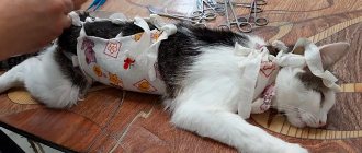
Traumatic injuries
The condition is accompanied by internal bleeding, which poses a threat to the animal’s life, so the cat should be urgently taken to the veterinary clinic.
Similar injuries can be sustained by an animal when it falls from a height.
A bruise or rupture of the spleen provokes a fall from a height, a blow to the stomach with a heavy blunt object. In addition to this organ, the liver, kidneys, brain, and limbs are injured, so injuries manifest themselves as a complex of symptoms. The most common:
- severe pain in the body, the animal meows loudly;
- lameness or deformation of paws;
- breathing, heart rhythm, swallowing disorders;
- vomit;
- urinating or passing bloody stool;
- refusal to eat;
- loss of consciousness.
Eosinophilic gastritis
Characterized by the accumulation of eosinophils in the gastric mucosa. It mainly affects older cats. The occurrence factors are low-quality food, allergies to food additives, uncontrolled use of medications by the cat by the owner, infection with worms. Manifestation:
- vomit;
- weight loss;
- loss of appetite;
- black color of stool due to coagulated blood.
Spleen tumors in dogs
Damage to the spleen is often observed with tumors of the hematopoietic system.
But the spleen as an organ itself can become the site of localization of primary tumors and metastasis of other malignant tumors. An enlarged spleen in dogs (splenomegaly) is found quite often. In 43-75% of cases, the cause is tumors.
What types of tumors and mass formations of the spleen occur in dogs?
1) Primary:
- - hemangioma
- - hemangiosarcoma
- - sarcoma (various types)
2) Secondary or multicentric:
- - lymphoproliferative or myeloproliferative diseases (for example, lymphoma)
- - malignant histicytosis, histiocytic sarcoma
- - hemangiosarcoma
- - mastocytoma
- - other malignant tumors with distant metastases (for example, melanoma).
3) Non-tumor causes of splenomegaly:
- - nodular hyperplasia
- - hematoma
- - thrombosis or heart attack
- - stagnant changes
- - extramedullary hematopoiesis
- - torsion of the pedicle of the spleen.
The most common tumor of the spleen in dogs is hemangiosarcoma. This is a high-grade tumor with hematogenous metastasis in the early stages of the disease. Rupture of the primary tumor can lead to acute and fatal hemorrhage. It develops in dogs aged 9-10 years.
What are the reasons for the development of spleen tumors?
The etiology of malignant tumors of the spleen is unknown. Their high prevalence among dogs of certain breeds (German shepherds, retrievers, Labradors) indicates the presence of genetic factors. Mutations of the PTEN gene may be involved in the mechanism of initiation and development of hemangiosarcoma.
What are the manifestations of splenic tumors?
Benign tumors of the spleen do not cause any clinical manifestations, even if they reach a significant size. The reason to consult a doctor is an increase in the volume of the abdomen, which occurs due to the growth of the tumor. Or such a tumor is discovered during a routine examination.
Animals with splenic sarcomas may develop nonspecific symptoms (eg, malaise). They are detected during examination, x-ray or ultrasound examination, and during diagnostic laparotomy.
Hemangiosarcoma may have the following manifestations:
- - apathy
- - weakness
- - pallor
- - anorexia
- - fainting
- - hemorrhagic diathesis (spontaneous recurrent bleeding and hemorrhages of varying duration and intensity)
- - heart rhythm disturbances.
And other more severe manifestations:
- — acute collapse after rupture of a primary space-occupying lesion
- - abdominal bleeding (into the abdominal cavity)
- - acute vascular insufficiency.
However, hemangiosarcoma may not give any clinical manifestations and may be an accidental finding by a veterinarian.
How to diagnose splenomegaly or splenic mass?
- General clinical blood test. Hemangiosarcoma gives a number of hematological abnormalities: anemia (decreased hemoglobin), acanthocytes (damaged red blood cells) and schistocytes (red blood cell fragments), thrombocytopenia (increased bleeding due to a decrease in the number of platelets).
- X-ray examination. Allows you to identify a tumor or fluid (in case of bleeding) in the abdominal cavity.
- Ultrasound. Allows you to obtain information about the structure of the neoplasm and its location in relation to normal spleen tissue.
- Needle biopsy (there is a risk of bleeding) - tissue is taken using a syringe with a thin needle and examined under a microscope.
- Excisional biopsy (diagnostic surgery involving removal of the entire tumor being examined). It is used if there is a clearly visible tumor in the spleen.
- To detect metastases, chest x-ray and ultrasound of other abdominal organs are used.
How to treat spleen tumors?
Treatment involves:
- Surgical removal of the tumor. Unfortunately, in the case of a malignant tumor, surgery does not provide a cure.
- Postoperative chemotherapy to prevent or delay the progression of micrometastases. Monotherapy or combination chemotherapy is carried out. However, survival is relatively short. For combination chemotherapy protocols, it is on the order of 141-179 days, and only less than 10% of dogs survive more than 1 year.
What's the forecast?
The prognosis for dogs with splenic hemangiosarcoma is poor. Metastasis in the early stages of the disease is typical for this type of tumor. In most cases, micrometastases are already present at the time the primary tumor is diagnosed. They progress rapidly and cause low survival - 15-86 days after tumor removal.
For other types of spleen sarcomas, the prognosis is also unfavorable. Survival is about 4 months. The cause of death of the animal is metastases.
Histiocytic sarcoma has an extremely unfavorable prognosis. Most animals are presented for euthanasia or die at the time of diagnosis from extensive metastases.
What are the consequences?
If splenomegaly is not treated, the following complications occur:
The pathology can result in a state of shock for the animal.
- Splenic rupture. When an organ is injured, a hematoma forms, which can rupture a few days after the injury. The condition threatens the animal's life, so the spleen is removed.
- Hemorrhagic shock. Appears with internal bleeding. The animal falls into a stupor, blood pressure drops, and death may occur.
- Cancer intoxication. Occurs due to the breakdown of tumor cells. The released toxins enter the bloodstream and spread throughout the body.
Spleen tumors in cats
Spleen tumors are less common in cats than in dogs. As in dogs, damage to the spleen is possible as a result of leukemia and lymphoma.
Classification of tumors and space-occupying formations of the spleen of cats
1) Primary tumors:
- - mastocytoma
- - hemangiosarcoma
- - sarcomas (various).
2) Secondary or multicentric tumors:
- - lymphoproliferative and myeloproliferative diseases (for example, lymphoma)
- - hemangiosarcoma
- - other malignant tumors with extensive metastasis (for example, adenocarcinoma).
3) Non-tumor causes of splenomegaly (or splenic masses):
- - nodular hyperplasia
- - hematoma
- - stagnant changes
- - extramedullary hematopoiesis.
About 15% of tumor pathologies of the spleen in cats are lymphoreticular and visceral mastocytomas.
Symptoms of visceral mastocytoma
- Malaise.
- Anorexia.
- Chronic vomiting.
Presumably, these symptoms are associated with the formation of ulcers in the stomach and duodenum due to the influence of histamine on H2 receptors in the stomach. As the disease progresses, ulcers perforate, peritonitis and death of the animal occurs. Cases of splenic rupture have been recorded.
- Anemia due to blood loss from gastric or duodenal ulcers (or as a result of bone marrow infiltration).
Treatment and prognosis of mastocytoma
Treatment consists of surgical removal of the tumor. The prognosis is unfavorable.
How is the treatment carried out?
Drug therapy
If the disease is caused by worms, then the pet is prescribed the appropriate drug.
Any drug must be prescribed by a veterinarian; self-medication is prohibited. Antibiotics are used for infectious diseases. To reduce the possible development of allergies, it is appropriate to prescribe antihistamines. To strengthen the body, treatment is supplemented with vitamin and mineral complexes. Chemotherapy is used for cancer. Parasites are expelled by anthelmintic drugs.
How should you prepare an animal for an ultrasound of the spleen?
The fasting diet for dogs is 8-10 hours, for cats 6 hours with free access to water. At the examination site, the hair is mechanically removed (with a clipper), and a special gel is applied. In emergency cases, the examination is carried out without preparation. The patient is placed on the right side or on the back. Ultrasound of the spleen is performed with 5 and 7.5 MHz sensors. Normally, on ultrasound, the spleen is elongated and homogeneous. The contour is smooth, clear, sometimes the capsule is visible. Vessels are visualized. In dogs it reaches a width of 3 cm, in cats 1.5 cm.
In veterinary clinics in Vitebsk, Mogilev, Novopolotsk, Minsk (Belarus) and St. Petersburg, Smolensk, Sevastopol (Russia) of the Veterinary Center of Dr. Bazylevsky A.A. You can get detailed advice on performing an ultrasound scan of the spleen, undergo an examination and receive the necessary recommendations for the treatment and prevention of spleen diseases.
Enlarged spleen in animals: how to treat
The spleen is a large formation that performs a large number of functions. If they are violated, various changes in this organ are possible, including an increase in its size. The development of splenomegaly in animals is a fairly common pathology associated with a large number of etiological factors. It is quite difficult to make such a diagnosis without special research, but its presence can be suspected by changes in the pet’s behavior. This is due to the fact that an enlarged spleen in dogs and cats causes certain unpleasant symptoms.