Eosinophilic granuloma is an inflammatory disease that cat owners often encounter. The exact cause of its development is unknown, and the pathological process is difficult to treat, so the sooner it is taken under control, the greater the chance of a complete recovery. What is eosinophilic granuloma in cats, and how is it treated?
Eosinophilic granuloma in cats
Eosinophilic granuloma in cats: description and causes
The mechanism of development of the pathology is associated with an increase in the level of eosinophils - cells that are responsible for the body's response to the effects of allergens and take part in the fight against pathogenic microorganisms. If their level increases significantly and remains high for a long period, the animal develops an inflammatory lesion of the skin and mucous membranes called granuloma.
With eosinophilic granuloma, the level of eosinophils in the body increases
The exact causes of the development of eosinophilic granuloma in cats are unknown, but most often the disease affects females aged 3 to 5 years. The risk group for the development of a pathological process includes animals with the following disorders:
- hypersensitivity of the body, which is expressed in frequent allergic reactions;
- chronic dermatitis and other skin diseases;
- genetic predisposition to the development of eosinophilic granuloma (if the disease was diagnosed in the mother, the likelihood of its occurrence in female kittens increases);
- hormonal imbalances due to sexually transmitted diseases, abuse of hormonal contraceptives, systemic diseases, etc.
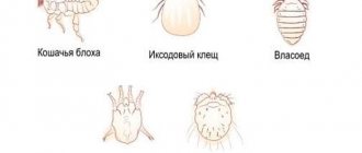
When allergens or any substances that provoke pathological reactions enter the body, an abnormal proliferation of cells begins in the deep layers of the mucous membranes and skin, which is called eosinophilic granuloma. The “trigger” mechanism for the development of the disease is infection with worms, flea and tick bites, and the presence of irritating factors in the food or environment.
For reference! Eosinophilic granuloma affects only representatives of the cat family, so there is no need to fear infection from contact with a cat. In humans, there is a disease with a similar name (it is also known as Taratynov’s disease), but it affects bone tissue, has a different nature and is in no way related to pathological processes in the body of cats.
Pet treatment
To accurately determine the diagnosis of granuloma in kittens, the veterinarian will examine the pet and, if necessary, conduct a microscopic examination (cytology). Based on the cytology results, the specialist will rule out other diseases with similar symptoms and perform a wet test to identify fleas and ticks. It often happens that it is impossible to determine the origin of the disease. Then complex treatment is applied. Local therapy for granuloma in cats is useless. Antiparasitic agents must be administered, regardless of whether the kitten has them or not. Only after this is it allowed to carry out anti-inflammatory therapy. Treatment is carried out mainly with the following steroid hormonal agents:
— Prednisolone; — Dexamethasone; - Triamcinolone.
Treatment is carried out for 21 days with tablets. If giving pills to a kitten is problematic, then you can replace them with injections. In this case, injections are given twice with a break of a week. Additionally, to reduce allergic symptoms, one of the following drugs is prescribed:
- Diphenhydramine; — Chlorambucil; - Cyclosporine.
In severe cases of the disease, a course of antibiotics is prescribed. After 1-1.5 weeks, the condition of a kitten with granuloma will improve significantly, but treatment cannot be interrupted. For older cats, therapy is increased if necessary to eight weeks.
Forms and varieties
Eosinophilic granuloma has several forms, and one cat can have one of them or several at once. All types of pathology have a similar cause, but different symptoms and features of the clinical course.
Table 1. Forms and features of eosinophilic granuloma.
| Types of disease | Features of the clinical course |
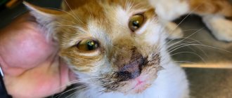
| Eosinophilic ulcer | The lesions are localized on the skin and mucous membranes around the oral cavity, as well as in the mouth, and can appear in cats of any breed and age, but in females they are three times more common. Their appearance is caused by an immune reaction to allergens and other irritating substances. Ulcers with a diameter of 2-5 mm have clearly defined edges, a red-brown tint, and other symptoms (itching, pain) are usually absent. |
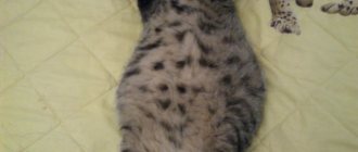
| Eosinophilic plaques | In the presence of predisposing factors, plaques can appear in all animals, regardless of age and breed. They are most often observed on the abdomen or the inside of the lower extremities, and can be single or multiple. Manifestations of this form of the disease look like oval red spots from 5 mm to 5 cm in diameter with well-defined edges, slightly raised above the surface of the skin. Plaques are often accompanied by ulceration, fluid exudate, and severe itching. |
| Allergic miliary dermatitis (eosinophilic plaque) | For the development of miliary dermatitis, a combination of provoking factors is necessary - for example, it often occurs in the presence of allergic reactions in cats infected with fleas or worms. The symptom is multiple rashes covered with crusts; in the chronic course of the disease, pigmented spots are observed. Another common sign of the disease is moderate to intense itching. |
| Eosinophilic granuloma | The main cause of the disease is allergic reactions of the body. It manifests itself as lesions of the mucous membranes and skin, which most often appear on the face, less often on the back of the thigh, but in general can be observed on any part of the body. Sometimes there is moderate itching and swelling of the tissues. |
For reference! The most common forms of eosinophilic granuloma are plaques and granulomas that appear on the paws and around the mouth.
What it is?
To summarize, we can say that this term refers to a wide group of inflammatory diseases of the skin of animals. There are various clinical forms, in many cases differing significantly in the nature of their course, but nevertheless an inflammatory reaction is almost always observed. Most often, the skin is affected on some part of the body, as well as the oral cavity (pictured).
Initially, it was believed that this disease could be initiated by dozens of reasons, including parasites, poor-quality food, stress and environmental influences, but today scientists are inclined to think that in almost all cases granuloma is a peculiar manifestation of a local and general allergic reaction. In most cases, skin lesions are very itchy, and therefore the animal constantly licks and scratches them.
Often this pathology is one way or another associated with flea dermatitis. Since owners for a long time do not attach any importance to the constant licking of their pet, the disease can go very far.
Symptoms
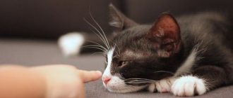
The symptoms of the pathology largely depend on its form, but all varieties have common signs. The first alarming symptom is the presence of skin lesions that look like round or oval tubercles with a shiny, sometimes moist surface. The hair around them falls out, and if there is itching, the pet will constantly itch and lick the affected areas. Sometimes their appearance is preceded by swelling, local edema and yellow-brown ulcers, which at first do not cause discomfort to the cat, but as the pathological process progresses they can grow and gradually spread to other parts of the body.
Typical localization of eosinophilic granuloma is the lower lip
Sometimes cats with eosinophilic granuloma become restless and show more aggression; when the lesions are localized to the paws, lameness appears, and if they grow inside the oral cavity and interfere with normal food intake, the symptoms include signs of exhaustion and dehydration.
Video - Skin diseases in cats and their diagnosis
Prevention
There is no specific prevention - a malfunction of the immune system can occur for reasons beyond the control of the owner.
General efforts are aimed at maintaining the health of the pet:
- proper feeding, maintenance, care;
- minimizing stress;
- annual preventive examinations;
- timely vaccination;
- treatment for internal and external parasites.
These same measures allow you to maintain stable remission. It is important to follow the prescribed diet, prevent parasites, and protect your chronically ill pet from stress. Months of treatment can be negated by a piece of chicken or an annoying guest with whom the cat clearly does not want, but is forced to contact.
Diagnostics
Diagnosis of eosinophilic granuloma consists of several stages and makes it possible to distinguish pathology from dermatological and oncological diseases with similar symptoms.
- Visual inspection. The doctor conducts a detailed examination of the skin lesions using special magnifying glasses and other devices.
- Anamnesis collection. Anamnesis collection is carried out in order to identify provoking factors that can cause an allergic reaction in the body. The owner is asked questions regarding the cat’s diet and living conditions, and he must talk in detail about all aspects of the pet’s life.
- Smear collection. After collecting an anamnesis and an external examination, the veterinarian takes a smear-imprint from the surface of the ulcers onto a glass slide, stains it with special substances and examines it under a microscope. The method allows you to determine the presence and type of pathogenic microorganisms that may be present on the surface of the skin, as well as detect eosinophils - a high concentration of these cells in the sample confirms the diagnosis.
- Blood tests. The study helps detect increased levels of eosinophils and other signs of the inflammatory process.
- Tests to identify parasites. They are carried out to identify fleas, worms and other parasites that can provoke pathological reactions in the body.
- Allergy tests. Intradermal tests to detect allergic reactions to various environmental factors.
- Biopsy. Tissue sampling is necessary when tumor processes are suspected, and is prescribed in cases where mast or giant cells are found in eosinophilic plaques, ulcers and papules.
To make a diagnosis, a comprehensive examination of the cat is necessary.
Attention! Treatment for a cat with eosinophilic granuloma can be prescribed only after a comprehensive diagnosis - the symptoms of the disease resemble manifestations of other pathological processes of an infectious, non-infectious or malignant nature.
How to treat a sick cat
Since the manifestations of eosinophilic granuloma are associated with systemic reactions of the body, and not with damage to the skin, the use of ointments, creams and other local remedies is not advisable. The basis of treatment is hormonal drugs:
- Dexamethasone (0.3 mg/kg per day));
- Prednisolone (2 mg/kg);
- Triamcinolone (0.8 mg/kg).
Steroids are taken in tablet form or administered subcutaneously, and the course of treatment should not exceed 3 weeks, and the exact dosage and frequency of use is determined by the doctor. As the skin lesions disappear, the dosage of the drugs is reduced and then discontinued completely.
Eosinophilic granuloma is treated with steroid hormones
Symptomatic medications (local drops and ointments that reduce itching), vitamins and antiparasitic agents are used as additional treatment.
In severe cases, when eosinophilic granuloma does not respond to treatment with steroids, immunosuppressants are used - drugs that inhibit the functioning of the immune system and reduce the intensity of its reaction to exposure to allergens.
Classification of immunobiological preparations
If formations on the skin and mucous membranes cause serious discomfort or interfere with normal eating, surgical intervention is performed - laser destruction, cryotherapy or excision of the affected tissue. Surgical treatment must be combined with conservative therapy, otherwise there is a high probability of disease relapse.
The presence of parasites is one of the factors provoking the disease
The prognosis of treatment depends on whether the provoking factor of the pathological process is identified and whether it is possible to eliminate it. If the animal's contact with the allergen is limited, eosinophilic granuloma is completely cured without complications or consequences.
Important! If left untreated, eosinophilic granuloma can develop into a malignant tumor, so at the first signs of pathology, the pet needs diagnosis and complex therapy.
Classification of immunosuppressants
Clinical case
Based on Lam S. et al, Eosinophilic granuloma/Langerhans cell histiocytosis: Pediatric neurosurgery update. 2015
A 17-year-old young man was hospitalized due to an increasing scalp lesion over the past 6 weeks. The formation is painful on palpation and periodically bleeds due to ulceration, but no neurological deficit has been identified. CT and MRI revealed a large lesion in the frontal bone on the right, compressing the superior sagittal sinus. A total resection of the formation was performed, and the diagnosis of Langerhans cell histiocytosis of the skull bone was confirmed. At the outpatient stage, cytostatic therapy was carried out.
Figure 6. (a) CT examination without contrast - frontal scan (upper and middle part) and 3D reconstruction of the skull (lower part). (b) MRI scan. T1-weighted image in the coronal plane (top) and T2-weighted image in the sagittal plane
Sources
- Coppes-Zantinga A., Egeler RM The Langerhans cell histiocytosis X files revealed. Br J Haematol, 2002. - V. 116 - N. 1 - P. 3–9.
- Lam et al., Management of adult patients with Langerhans cell histiocytosis: recommendations from an expert panel on behalf of Euro-Histio-Net. Orphanet J Rare Dis, 2013 - V. 8. - N 72.
- Lam S., Reddy GD, Mayer R., Lin Y., Jea A. Eosinophilic granuloma/Langerhans cell histiocytosis: Pediatric neurosurgery update. Surg Neurol Int, 2015. - N. 6 (Suppl 17): S435 - S439.
- Langerhans' Cell Histiocytosis (Histiocytosis X). What is it? Harvard Medical School. Harvard Health Publishing. October, 2014. www.health.harvard.edu
- Sharma R., Singh R. et al. Langerhans cell histiocytosis (skeletal manifestations). Radiopaedia. https://radiopaedia.org/articles/langerhans-cell-histiocytosis-skeletal-manifestations-1
- Shea C. R, James W. D. et al. Langerhans Cell Histiocytosis. Medscape, 2021. https://emedicine.medscape.com/article/1100579‑overview
- Churilov L.P. Death on takeoff, or Who are you, Doctor Taratynov? Health is the basis of human potential: problems and ways to solve them, 2014. - T. 9 - No. 2 - P. 919–929.
- Yusupova L. A., Yunusova E. I., Garayeva Z. Sh., Mavlyutova G. I. Histiocytosis X. Practical Medicine, 2014 - T. 08 - No. 14.