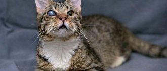
Cats develop an eyesore quite often. Older animals are most susceptible to this unpleasant symptom. In veterinary practice, blurred vision is called leukoma. This pathology can seriously worsen the pet’s quality of life and cause a number of complications, so it is important to begin its therapy immediately after diagnosis.
Causes
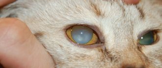
The external manifestation of an eyesore in a cat can be confused with a cataract, but during leukoma it is the cornea that suffers, not the lens. The described pathology is mainly diagnosed due to eye damage due to ulcers, as well as various types of keratitis. However, there are other factors that can provoke the development of eyesores:
- corneal damage, trauma, infection and inflammatory processes;
- chlamydial trachoma;
- accumulation of leukocytes and proteins in the fluid inside the eyes;
- vitreous diseases.
Prevention
Preventive measures include preventing eye injuries, promptly consulting a doctor at the first signs of ophthalmological diseases that can lead to the formation of a cataract.
You can undergo a comprehensive examination, biomicroscopy and receive surgical treatment for a cataract at the Optic-Center ophthalmology clinic.
Sign up
for an initial consultation with an ophthalmologist at the Optic Center clinic by calling
8-800-775-78-58
or on our website
Sign up for a comprehensive examination
Classification of pathology
A thorn is usually divided into congenital and acquired. If such a spot appears on a kitten immediately after birth, then veterinarians believe that during pregnancy the female suffered some kind of inflammatory or bacterial illness. The acquired disease is observed due to the factors described above. The disease is classified as follows depending on its location:
It is possible for the disease to develop with only the pupil of the eye affected.
- Total view. The thorn is observed all over the eye.
- Central. Only the pupil is noticeable.
- Peripheral. A certain part of the visual organ is clouded, bypassing the pupil.
First aid
Self-medication in this case is strictly prohibited. Medicines are used only after consultation with a veterinarian. If you delay in going to the clinic, your pet’s condition will quickly deteriorate.
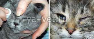
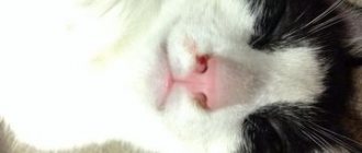
As soon as the owner notices that his pet has a white film on the organ of vision, and an urgent visit to the veterinarian is impossible, the animal is treated at home.
Wiping the eyelids
Take a clean cotton cloth, moisten it with warm filtered water, and carefully remove all discharge from the eyes from the outer corner to the inner one.
Moisturizing the eyes
If the eye is swollen, carefully spread the eyelids and drip saline solution. If the film falls out, periodically moisten the eye with saline solution and apply a sterile bandage.
Help with bleeding
If bleeding occurs, apply a sterile bandage or gauze folded in several layers to the eye (the material should not stick). When soiled, the bandage is changed, but at least once a day.
Examination by a veterinarian
At the clinic, the doctor conducts a full examination of the pet. An ophthalmoscope is used to examine the condition of the fundus.
If there are no injuries, clinical tests are prescribed.
What symptoms indicate illness?
The first sign that indicates the occurrence of a cataract is cloudiness of the organ of vision. However, there are other symptoms that help to suspect the presence of leukoma. Often, in the initial stages of development, cats begin to have difficulty navigating in a room that was previously very familiar to them. Animals bump into objects, owners. The following symptoms are also identified:
- nervousness and anxiety;
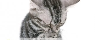
- hissing when washing or touching the owner's visual organs;
- development of strabismus;
- redness of the mucous membrane of the visual organs;
- swelling of the cornea and increased production of tears.
If a peripheral or central type of disease is observed, the cat does not always lose visual functions; sometimes they are preserved. The total type of pathology dooms to complete loss of vision.
Structure and functions of the cornea of the eye
The cornea looks like a concave-convex natural lens. This section of the eye is up to 10 mm in diameter.
The structure of the cornea is represented by 5 layers:
- anterior (integumentary)
, consisting of epithelial cells, providing protection to this part of the eye from damage; - Bowman's membrane
, which helps the cornea maintain its shape; - stroma
, formed by collagen fibers and leukocytes, making the cornea of the eyes quite strong; - Descemet's
, which increases the cornea's resistance to harmful factors; - posterior (endothelial)
, supplying the cornea with nutrients.
The cornea performs several important functions - it participates in the process of light refraction, protects the eye from injury, and maintains the normal shape of the eyeball. If its transparency deteriorates, a sharp deterioration in vision occurs, requiring immediate eye treatment.
Diagnostic measures
With such a pathology, the animal must undergo gonioscopy.
If owners notice that a cat has an eyesore, it is important to contact a veterinarian as soon as possible. The doctor will conduct a survey during which he will find out what the animal is fed and what pathologies it has suffered. Taking an anamnesis helps to identify the cause that provoked the occurrence of a cataract. Then the doctor begins a visual examination of the pet’s visual organs and conducts tonometry, which makes it possible to detect intraocular pressure to confirm or refute the presence of cataracts and glaucoma. The cat is also sent for the following examinations:
- biochemical and general blood tests;
- serology;
- fluorescein test;
- gonioscopy.
Recommendations for owners
After treatment, the veterinarian will provide recommendations to cat owners on care and nutrition, which owners must strictly follow, constantly monitoring the condition of the eyes and health until the animal is completely healed.
For eye rinsing and hygiene, medicinal decoctions of medicinal herbs and ophthalmic solutions (boric acid, warm water, decoction of chamomile, sage) are prescribed. Potassium permanganate lotions, tricylan powder, and furatsilin solution (the tablet is diluted in water) also help well.
Advice! After therapeutic therapy, even if the cat’s white film has disappeared from his eyes, his condition has improved, do not be lazy to take your pet to the clinic for a second scheduled examination!
What to do?
Treatment with medications
If the pathology is diagnosed at the initial stage of development, it is treated with medications that make it possible to eliminate the source of infection and pain in the animal. Therapy should be prescribed as quickly as possible so that a scar does not form on the visual organ. To remove the cataract, veterinarians from the TsNVOiM clinic recommend using Levomycetin or Dexamethasone. Injections using Novocaine, as well as boric acid in the form of a solution, can also be prescribed. Tetracycline ointment and Actovegin, which is available in gel form, will help cure the thorn.
Surgical intervention
If the animal’s disease has reached an advanced stage, then it requires surgical treatment.
The operation is prescribed in an advanced stage, as well as in a situation where drug treatment does not bring the required therapeutic effect. Keratoplasty is often prescribed, during which the cornea is transplanted. Sometimes tarsography is also prescribed, during which the surgeon sutures the edges of the eyelids. This procedure can be either complete or partial. With the help of this surgical intervention, it is possible to protect the cornea, as a result of which the cat recovers quickly.
ethnoscience
It is permissible to resort to the help of healers’ recipes exclusively in combination with traditional methods of therapy. A solution containing honey will help treat a cat’s eyesore. You will need to add a small amount of the ingredient to the water and mix thoroughly, achieving a liquid consistency. Powdered sugar can be used instead of honey. You will need to instill the resulting composition 3 drops into the affected visual organ daily until you can completely get rid of the pathology. It should be noted that treatment with this method will be ineffective if the cat’s cataract is caused by an infectious lesion.
Treatment of a cataract
Drug therapy in the treatment of cataracts is carried out in the case of residual inflammatory processes occurring in the cornea. In this case, the choice of medications is directly related to the cause of the disease - these are anti-tuberculosis or antiviral medications, which in rare cases lead to a slight clearing of leukoma. However, drug treatment cannot restore visual acuity and does not completely cure the cataract.
Experts consider a modern effective way to radically get rid of a cataract to perform a surgical operation - keratoplasty. As a result of a rather complex surgical intervention, the damaged area of the cornea with a cataract is replaced with a completely transparent graft. During the operation, donor tissue or a special corneal transplant produced using special technology, like a medical product, can be used.
The operation is performed on an outpatient basis, using local anesthesia or general anesthesia.
The rehabilitation period after keratoplasty surgery can take quite a long time - up to a year inclusive. This is due to the individual characteristics of the patient’s cornea, his age and state of health. During the entire rehabilitation period, be sure to regularly (according to the established schedule) visit an ophthalmologist.
The prognosis for keratoplasty is favorable in most cases.
Rehabilitation after therapy
During the recovery stage, the animal must receive high-quality and healthy nutrition.
Treatment usually takes a long time, on average about 3 months. After therapy and elimination of eye cloudiness, animals are often prescribed antiviral medications and deworming. In addition, it is important for the owner to monitor the animal’s diet. It is permissible to use only high-quality complementary foods recommended by the veterinarian. If surgery has been performed, immediately after it you should slightly limit your pet’s physical activity, and also regularly visit a veterinarian to reduce the likelihood of developing postoperative complications.
Diseases of the cornea of the eye
There are various eye diseases that affect the cornea. Main pathologies of the cornea:
- keratitis;
- xerophthalmia;
- keratoconus;
- keratomalacia;
- bullous keratopathy;
- corneal dystrophy.
In addition to the listed pathologies, the cornea can be subjected to mechanical, thermal effects, and damaged by foreign bodies entering the eye.
Keratitis
The disease leads to inflammation and clouding of the cornea, the appearance of ulcers and pain. Other symptoms of eye pathology:
- lacrimation;
- photophobia;
- redness of the eye;
- decreased visual acuity.
Keratitis can be infectious (viral, bacterial) or non-infectious (occurring after injuries to the cornea or the entire eye, against the background of allergies, diabetes). In the absence of adequate treatment, the disease inevitably causes complications, among which the most dangerous are cataract and blindness.
Xerophthalmia
With xerophthalmia, drying of the cornea and conjunctiva of the eye occurs. The disease occurs with unpleasant symptoms:
- burning, itching in the eye;
- photophobia;
- swelling of the eyelids;
- increased eye fatigue.
There are many reasons for the development of xerophthalmia. These may be chemical burns of the cornea, chlamydia damage, menopause and menopause, improper use of contact lenses for the eyes, increased visual stress. Treatment of the cornea most often takes a long time and requires an integrated approach.
Keratoconus
The disorder is degenerative in nature. As it develops, the cornea of the eye begins to thin and takes on the shape of a cone. The pathology is accompanied by progressive deterioration of vision, diplopia (double image). In severe cases, Descemet's membrane ruptures and the cornea swells.
This disease can occur after damage to the eyeball. Other reasons for the development of ocular keratoconus are disorders in the endocrine system and negative heredity. In many cases, the pathology is combined with other diseases. It can affect the cornea of the eye against the background of eczema, asthma, hay fever, so the patient needs especially careful treatment.
Keratomalacia
With such an anomaly, foci of necrosis appear on the cornea. At the same time, other parts of the eye (conjunctiva, lacrimal glands) may be affected. Keratomalacia occurs with swelling of the cornea, severe lacrimation, increased sensitivity of the eyes to light, and deterioration of visual perception.
The main factors in the development of corneal disease are protein starvation and vitamin A deficiency. Eye keratomalacia can accompany liver diseases (viral hepatitis, cirrhosis). Treatment of corneal pathology is carried out comprehensively.
Bullous keratopathy
If bullous keratopathy develops, fluid accumulates in the cornea of the eye. This leads to the formation of abscesses (bulls). They burst, cause pain, and lead to clouding and swelling of the cornea.
The main causes of corneal pathology:
- eye injuries;
- infections;
- Fuchs' dystrophy;
- poor-quality eye surgeries.
This corneal disease is considered dangerous because it can cause irreversible vision loss. The treatment regimen for bullous keratopathy is selected taking into account the stage of the disease and the severity of negative changes in the eye.
Dystrophic pathologies of the cornea
Corneal dystrophies are hereditary diseases that affect both eyes. Symptoms of such pathologies:
- clouding of the cornea of the eye;
- photophobia;
- lacrimation;
- feeling of a foreign object in the eye;
- deterioration of visual function.
There are different forms of corneal dystrophy - stromal opacification, band-like, endothelial, marginal degeneration. Eye disease is rapidly becoming severe and therefore requires timely detection and effective treatment.
Preventive actions
To prevent the cat from developing an eyesore, you should systematically examine the animal’s visual organs. It is important to normalize your pet’s diet by adding a sufficient amount of vitamins and microelements. In addition, veterinarians recommend giving special cat vitamins from time to time. It is important for owners to take care of creating a safe environment for their cat to live in and protect it from various injuries. If the pathology has already been diagnosed, it is important to start treatment immediately and follow all the veterinarian’s recommendations. It is recommended to place the animal in a separate room where there are no sharp objects that it can hit.
If you have leukoma on the visual organs, you should not let your pet outside unattended. It is also important to keep it away from stray animals, as they may be infected. It is forbidden to carry out independent treatment and use for a cat the same medications that were prescribed to a person. This precaution is due to the fact that medications may not only not cope with the disease, but also cause serious adverse reactions in the cat.
Alarming symptoms
When a white-gray film appears on the eyes of a furry pet, owners should carefully examine the eyes of the pet and pay attention to the general condition and behavior of the cat.
Alarming symptoms:
- frequent blinking;
- tearfulness;
- discharge from the eyes of a different nature;
- changes in behavior (lethargy, inactivity, restlessness);
- loss of appetite, digestive problems;
- fever, chills;
- change in the shade of the mucous membrane;
- swelling of the eyelids;
- lack of pupil reaction to light;
- photophobia.
With this problem, cats prefer to retire to secluded places and are reluctant to make contact. Restless behavior gives way to apathy and depression. The pet constantly rubs its eyes with its paw, shakes its head, blinks frequently, squints its eyes, and cannot tolerate bright light. Purulent, serous exudate may accumulate in the corners of the eyes.