Treatment of fractures in cats is one of the main surgical areas in veterinary medicine.
A fracture is a complete or partial disruption of the integrity of a bone, which occurs due to an external mechanical force that exceeds its strength. Fractures in cats can be classified into two groups: traumatic and pathological. Traumatic fractures occur due to mechanical impact, in cats most often as a result of a fall from a height or auto injuries, as well as bites by dogs. Occasionally, we also receive cats with gunshot fractures.
Pathological fractures in cats occur even with a small impact, as they say, “out of the blue.” They occur when the bone becomes more fragile as a result of some pathological process - usually this is how bone tumors or metabolic disorders associated with poor nutrition affect the bone tissue. In cats, such problems are usually associated with so-called secondary nutritional hyperparathyroidism. Under this terrible name lies a banal reason - with an excessive meat diet, too much phosphorus and very little calcium are supplied with food. As a result, calcium for the body's functioning - maintaining muscle and heart function - is washed out of the bones under the influence of parathyroid hormone. Bones become very fragile and break “out of nowhere.”
Doctors divide fractures in cats into closed (the integrity of the skin or mucous membrane is preserved) and open (the skin or mucous membrane, for example, in the oral cavity, is broken by a bone fragment); into simple (the bone breaks into only two parts) and complex (comminuted fractures); into transverse, oblique and helical – according to the shape of the fracture; intra-articular fractures (those that occurred in the intra-articular cavity) are also distinguished.
What symptoms may indicate that a cat has a fracture?
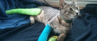
If a cat has a broken paw (front or back) - and veterinarians have to treat such fractures most often, you will see the following signs of a fracture:
- the animal does not lean on the affected limb at all
- the cat is taking care of its sore paw, an attempt to touch it results in a flash of pain, your pet may try to bite you
- severe swelling of the soft tissue appears at the site of the fracture - the diseased paw is twice as thick as a healthy one, a large hematoma may appear
- the configuration of the diseased paw is changed, it seems asymmetrical in relation to the healthy one
- a bone fragment can be seen through the skin
With specific fractures in cats, you can notice other external signs - for example, if the cat has a broken jaw, it cannot eat, trying to open its mouth causes pain, the muzzle may lose symmetry. If a cat has a fracture of the spine or pelvis, she cannot rest on her hind legs, they drag, and control over bowel movements and urine output is often lost.
Types of osteosynthesis
Today, bone fusion in small domestic animals using fixing metal structures can be carried out according to different schemes. The choice of surgical technique and fixation of bone fragments is determined primarily by the nature of the damage:
- Osteosynthesis of the femur in cats is most often performed using the intramedullary method. A knitting needle or pin is placed inside the bone, which is used as a basis for aligning bone fragments.
- The bone surgery is performed differently: the fixing elements are not inserted into the bone cavity, but are applied to it and secured with screws or other fasteners. Using this scheme, osteosynthesis of the radius bone in cats or fixation of other flat bones can be performed.
Note! The cost of osteosynthesis surgery in cats using extraosseous elements is almost always higher than when using intramedullary wires and pins. The choice to install overlay plates is made primarily when it is impossible to fix the damaged area in any other way.
- Extrafocal surgery can be performed not only for a fracture, but also for a dislocation. Above and below the site of injury, wires are inserted into the bone and fixed from the outside with a polymer compound. This technique is considered as an alternative to external fixation of plates, primarily due to its lower cost.
Here we should also consider osteosynthesis in cats using Kirschner wires and the practice of using the Kirschner apparatus. Knitting osteosynthesis operations in cats are carried out quite often, but the use of an external apparatus is limited due to the small size of the animal. However, this problem can be solved - the knitting needles are not fastened with mechanical clamps during installation, but are fixed using high-adhesive adhesives (analogues of “cold welding”).
What can you do yourself to help your cat?
The main task of first aid for a fracture in a cat is to understand whether there is bleeding and stop it. As a rule, severe bleeding accompanies open fractures, fractures caused by bites and gunshot fractures. To stop bleeding, it is better to use a pressure bandage, which is effective in 90% of cases. We recommend a pack of sterile gauze pads to stop bleeding. If they are not nearby, then you can use a handkerchief, mitten, piece of fabric, or feminine pad. Sterility is not so important now, the main thing is to stop the bleeding, which is life-threatening for the cat. A swab made of napkins or other fabric should be placed directly over the source of bleeding and bandaged quite tightly - with gauze or an elastic bandage, or a piece of fabric. Now - hurry up to the veterinary clinic. Our doctors recommend not wasting time calling a doctor to your home in such a situation, because full care for a fracture can only be provided to a cat in a clinic setting.
If there is no bleeding, but you see that the cat most likely has a broken front or back leg and it is “dangling around a lot,” you can immobilize it in the position it is in. Don’t try to “set” the fracture yourself! This is very painful. If handled improperly, the sharp edges of the bone can damage blood vessels and nerves, causing pain and additional injury to the cat. You just need to ensure that the cat's paw remains motionless while transporting the animal to the clinic. If you are in doubt or afraid, it is better not to do anything and take your cat to a veterinary clinic as quickly as possible.
First aid
If a fracture of bone structures is suspected, the animal must first be provided with complete rest. The injured cat must be moved to a flat surface. A wide board, a plastic panel from a car, thick cardboard or plywood are suitable for this purpose. If you don’t have such an object at hand, you can wrap the cat in a towel or blanket.
Transporting a cat with a pelvic fracture
To ensure immobilization, the pet should be secured to a hard surface using ropes or belts. In field conditions, a belt, harness, or cord are suitable. The cat must be secured on the board in the forearm area. An injured animal should be transported in the back seat of a car. It is important that damaged bones are kept at rest.
Despite the severe pain, veterinarians do not recommend giving your pet any painkillers. In the case of an open fracture, the owner must take measures to stop the bleeding, cover the wound with a clean cloth and transport the animal to a specialized facility as quickly as possible.
How will the doctor act?
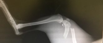
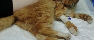
First, the doctor will numb your animal. He then examines the cat and its broken leg. This is of particular importance for those patients who were injured by a car or fell from a height, because in addition to the problems that are obvious to the owner - a fracture - the cat may have a chest or abdominal injury. These injuries to a cat may not be noticeable, but require significantly more urgent and complex treatment than a fracture. After the examination, your veterinarian will take an X-ray of your cat's broken leg to evaluate the fracture and plan treatment, as well as further diagnose other injuries, if any. Sometimes sedation is required for x-rays.
As a rule, 99% of fractures of the front and hind legs in cats require treatment with surgery - osteosynthesis. This operation is performed as planned, often 3-5 days after the fracture. This period is explained by the physiological characteristics of the body: during an injury, massive hemorrhage occurs in the area of the fracture, and then this blood and parts of the destroyed tissue that fall into it become so-called “osteogenic elements” - substances that stimulate the bone to heal. If surgery is performed immediately after a fracture, the entire contents of the hematoma will spill out, and healing will be slower and more difficult. Another difficulty for manipulating bone fragments is created by swelling of the soft tissues, which subsides precisely by 3-5 days after the fracture. The exception is open fractures - due to the open gate for infection, they require urgent (within 24 hours) surgery.
Before surgery, the doctor will fix the broken paw with a bandage.
Of course, fractures of the jaw, pelvis, and spine require a special approach in cats—we’ll talk about them a little later.
Clinical manifestations
Once it occurs, the pain syndrome provoked by metastases is moderate in nature. It intensifies after physical activity or at night.
As the tumor process develops, the symptoms also intensify. The pain syndrome becomes pronounced and painful.
There are other “signals” indicating the presence of bone metastases:
- Spinal cord compression. It happens quite rarely, but it makes life quite difficult. The thoracic spine or the lumbar and cervical sections are subject to compression.
- Hypercalcemia. The level of calcium in the blood increases, which ultimately leads to a malfunction of many body systems. The complication is very common.
- Pathological fractures. The femur in the area of the diaphysis or neck often suffers.
Patients with late stages of cancer also exhibit symptoms such as:
- Temperature increase;
- Exhaustion;
- Weakness, etc.
Why is osteosynthesis necessary when you can simply apply a cast?
Plaster casting is not used to treat fractures in cats for many reasons. To begin with, it is very difficult, even impossible, to force a cat to take care of a broken paw with a cast. And, even more so, provide the animal with bed rest or hang its leg in traction. Cats try to get rid of the cast as quickly as possible, bite it, gnaw it, try to remove it on furniture, causing additional harm to themselves. The second reason is that since cats and dogs almost never break their legs by “slipping on ice” or “unsuccessfully jumping,” they almost never have fractures of the “crack” type or simple fractures without displacement. For this reason, fractures in cats often require complex reduction of fragments and comparison of fragments. Finally, a number of studies have shown that for the fastest and most complete healing of a fracture, several factors are required - the most complete comparison of fragments, their most rigid fixation, preservation of blood supply and early support on the limb. All these requirements cannot be met if fractures of paws in cats are treated with the application of plaster, therefore, throughout the world, osteosynthesis is preferred in both veterinary and human orthopedics, in which recovery occurs faster and with better quality.
What operations are there?
Osteosynthesis is performed using metal structures with which parts of the bone are fixed to each other. Metal parts can be placed inside the bone (pins, wires), pass through the bone (screws, screws, wire sutures) or attached to its surface (plates). In addition, there are methods for fixing fractures in which the wires pass through the bone fragments, and the main structure that ensures the strength of their connection is located outside the limb (Ilizarov apparatus and other external fixators).
Osteosynthesis is performed under general anesthesia. The doctor selects a fixation structure depending on the type of fracture and accompanying soft tissue injuries. In veterinary medicine, we specialize in complex fractures (comminuted, crushed, multifocal, gunshot). We have all the necessary equipment and experience to treat them, including using external fixators and the Ilizarov apparatus. Many cats, who in other clinics were offered amputation of their paws due to complex fractures, retained their limbs thanks to the joint efforts of our doctors and their owners.
Improved technique
The Ladisten Orthopedics and Traumatology Clinic offers an alternative solution. Its founder, Dr. Veklich, improved the Ilizarov apparatus. The Veklich apparatus is installed in a minimally invasive way. It can be used to increase height or correct disproportion of limbs.
In case of bone injuries, surgical intervention to install the device cannot be avoided. Its design is almost identical to the Ilizarov apparatus, but instead of spokes, titanium rods with a diameter of 6 mm are used. The rod can be applied only on one side, reducing the risk of osteomyelitis significantly. It also does not come into contact with nerves and blood vessels. The entire structure is not too bulky and reliably holds damaged bone fragments.
How is the postoperative period going?
As a rule, after osteosynthesis, the cat remains in the hospital for 6 hours to 24 hours under the supervision of doctors. Then, suture treatment and painkillers are usually prescribed for 3-5 days. We recommend restricting movement for 4-6 weeks (cats can be placed in a large cage). The cat usually begins to lean on its paw 3-5 days after surgery.
After 3-4 weeks, you will need to visit the veterinary clinic with your cat again to take a control x-ray to assess the speed of fusion. On average, the rate of fusion, depending on the complexity of the fracture, varies from 2 to 8 months. After fusion, with the exception of rare cases (intra-articular fractures, fractures of the pelvic bones, very complex fractures in which the fixing elements firmly grow into the general mass of callus), metal structures are removed.
Primary tumors
Primary occurrence of bone cancer is rare. Among such diseases are sarcomas. The primary lesion can be caused by active growth of bone tissue, this occurs in people under the age of twenty.
Symptoms of the primary disease:
- Pain in the focal area. At first, the pain syndrome is mild or moderate. He may disappear, but then appears again. At later stages it is constant and pronounced.
- General symptoms that are characteristic of any cancer disease, for example, weight loss, weakness, etc.
- Manifestations characteristic of bone damage - lameness, pathological fractures, etc.
Primary and secondary diseases are united by a number of manifestations, which is why in making a diagnosis you should not rely only on the patient’s complaints. Diagnostics required.
What are the characteristics of different types of fractures?
Previously, we looked at the actions of cat owners and veterinarians in the event of a “paw fracture” - that is, a fracture of the long tubular bones of the limbs. In a cat, these are the following types of fractures with their symptoms and treatment: hip fractures - that is, the femur, as well as the neck and head of the femur, tibia fractures - the tibia and fibula, humerus fractures - the humerus and forearm fractures - the radius and ulna. . These types of fractures are encountered most often in the practice of a veterinary traumatologist. Let us now move on to the nuances characteristic of other types of fractures.
Plaster casts or internal fixation?
Bone osteosynthesis in cats is carried out for fractures and involves:
- Surgical intervention with the combination of fragments of damaged bone.
- Fixation of bone fragments with metal devices (screws, knitting needles, plates) until complete fusion, followed by removal of the fixing elements.
Despite the fact that osteosynthesis is a painful operation for cats and very labor-intensive for a veterinarian, there is currently no alternative to it. Self-healing, that is, healing of a fracture without medical intervention, is rarely 100% successful and almost always causes either complications or partial loss of functionality of the damaged limb. Attempts at fixation using plaster casts or splints also do not demonstrate the desired effectiveness: complete immobilization of damaged bones cannot be achieved due to the high activity of the animal.
Despite the fact that the price of osteosynthesis in cats can be significant, we recommend focusing on this particular treatment strategy due to its effectiveness and ease of the rehabilitation period.
Fractured toes in a cat
Owners usually refer to fractures of all the “small” bones of the hand and foot in cats as a “finger fracture.” Thus, these are fractures of the bones of the wrist and tarsus, and fractures of the metacarpus and metatarsus, and fractures of the small bones that make up the fingers. Cats often get these types of fractures after falling out of a window; they can happen if the cat's paw is stepped on. Sometimes such fractures are the result of a car injury.
External signs of these fractures are either a complete inability to step on the paw, or very severe lameness; Toe fractures in cats are not often open and are rarely accompanied by severe swelling. However, there is usually a significant pain reaction.
Surgical treatment (osteosynthesis) is necessary in case of fractures of the wrist and tarsus bones, less often used for fractures of the metacarpal and metatarsal bones (more often if all bones are broken), and is extremely rarely used for fractures of the finger bones - as a rule, fixation with a bandage and restriction of motor activity is sufficient for 1-1.5 months.
In any case, you need to consult a veterinarian and x-ray the limb.
Consequences that may arise
One of the common complications of pelvic bone fracture is improper healing of damaged structures. In this regard, the animal may experience lameness, impaired motor activity, and changes in gait.
With concomitant injuries and complex fractures in domestic cats, pelvic canal stenosis is often observed. The complication develops over time, represents a narrowing of the pelvic ring and is accompanied by difficulties with bowel movements and chronic constipation.
In case of damage to the nerve endings, the cat often experiences loss of sensation in the hind limbs, back and tail.
We recommend reading about a cat's tail fracture. You will learn about the causes of injury, signs and symptoms, diagnosis, and treatment options. And here is more information about what to do if a cat is lame on its paw.
Spinal fracture in cats
This type of fracture is one of the most difficult in veterinary practice. What are the symptoms of a spinal fracture in cats? A spinal fracture in a cat is the result of a serious injury - a car injury, a fall from a height, serious bites and bruises. As a rule, these fractures occur in the thoracic or lumbar spine, then the cat cannot rest on its hind legs (paralysis of the hind legs), and sometimes urine flows from it. If a cat has damaged the spine in the sacral region, the ability to walk is retained, but there are difficulties with urination/defecation. If a cat breaks its spine in the neck area, it can be completely paralyzed - both its front and hind legs. These injuries are extremely painful and your cat may bite or scratch you when you touch them. Cats with such injuries must be moved extremely carefully; a rigid stretcher is preferable. But if there is no hard one nearby, don’t waste time looking. Bring your cat to the veterinary clinic as soon as possible. In such cases, the clock counts, so don’t hesitate. We categorically do not recommend calling a doctor to your home in such cases - he will only be able to anesthetize the animal, in the case of a spinal fracture, this will only be a waste of time.
The fact is that when a cat has a spinal fracture, the biggest problem is damage to the spinal cord from fragments. This is the cause of paralysis. Nerve fibers can be torn - and then, unfortunately, the situation is irreversible. Or fragments and splinters may simply compress the spinal cord. In this situation, the sooner help is provided to the animal, the greater the chance of saving it and restoring normal support.
Treatment of a spinal fracture in a cat, cat or kitten is always surgery. During this process, the doctor will examine the spinal cord and determine whether it is intact and whether there is hope for restoration of all functions. Then he will remove small fragments and fix the damaged vertebrae in their normal position - usually knitting needles and screws are used for this. After such an operation, the cat usually remains under observation in the hospital from a day to a week, depending on its condition. If the spinal cord is not severed, recovery usually occurs 3-4 weeks after surgery, and the first signs of improvement appear the very next day after surgery.
Veterinary clinic of Dr. Shubin
Introduction
The main cause of pelvic bone fractures is trauma; most often, these fractures are caused by a road traffic accident, but any blunt trauma can be accompanied by damage to the ilium, ischium and pubic bones.
Fractures can occur on the wing of the ilium, the body of the ilium, the body of the ischium and the body of the pubis (and more often the pubic symphysis). The pelvis itself is a box-shaped structure, and for one segment to be displaced by a local fracture, three more fractures must be present in other places. In most cases, the iliac, ischial and pubic bones are broken at the same time. A fracture of the ilium is of particular importance; it is this bone that transfers the physical load from the hind limb to the body of the animal (through the sacrum and spinal column). In addition to impaired support function, a fracture of the ilium is usually accompanied by a narrowing of the pelvic ring, which can subsequently lead to problems with feces and labor disturbances. The most common form of ilium fracture is a long oblique fracture of the body of this bone, but transverse and comminuted fractures are also likely to develop. When the ilium is fractured, the caudal fragment is usually displaced medially and cranially, which narrows the lumen of the pelvic canal.
Isolated fractures of the ischium and pubis are rare, but when associated with other pelvic fractures, reduction and stabilization of the primary weight-bearing segments usually leads to acceptable reduction and stabilization of the ischium and pubis. The most common indication for surgical intervention for fractures of the pubic and ischial bones is soft tissue hernia. Hernias of the serous membrane of the abdominal wall can be caused by separation of the pubic symphysis or avulsion of the cranial pubic ligament. Rarely, hernias may develop caudal to the acetabulum.
A pelvic fracture in dogs and cats is a severe injury caused by significant physical forces, and associated soft tissue injuries are often observed. Of primary importance are injuries to the lower urinary tract (ruptures of the bladder and urethra), muscle avulsions at the site of attachment of the rectus abdominis muscle and the formation of a hernia of the serous membrane of the abdominal cavity, damage to the intestines of the pelvic canal, as well as damage to the lumbosacral plexus or sciatic nerve. When examining an animal with pelvic fractures, it is always important to keep in mind the possibility of associated soft tissue injuries and promptly evaluate them (eg, complete neurological examination, thorough physical examination with rectal palpation, contrast radiographic examination of the lower urinary tract).
Clinical signs and diagnosis
As mentioned above, the main cause of pelvic bone fractures is trauma, therefore, there is no species, gender or age predisposition for pelvic bone fractures. More often than others, this type of fracture can be recorded in animals with access to the street.
A physical examination of the animal reveals lameness with a lack of support function; with minimal displacement of fragments and mild damage to soft tissues, a partially preserved support function of the affected side may be noted. Bruises often form in the area of pelvic fractures; if there are bruises on the ventral surface of the abdomen, urethral injury should be suspected. During examination, special attention is paid to the integrity of the abdominal muscles, sciatic nerve function and rectal palpation.
The main method for diagnosing pelvic fractures is radiographic examination; two orthogonal projections are usually required (ventrodorsal and lateral). Due to the severe pain associated with these fractures, sedation or general anesthesia is necessary to adequately position the animal on the table. When assessing radiographic images, differentiation is made between sacroiliac fracture, acetabular fracture and hip dislocation.
Additional tests (eg cystography, urethrography) may be required to identify soft tissue damage.
When examining blood, there are no signs characteristic of a pelvic fracture; this type of laboratory analysis is usually used to identify concomitant diseases that can increase anesthetic risks. Also, before planned surgical treatment, a thorough assessment of the animal is carried out; diagnostic tests may include radiographic examination of the chest and abdominal cavities, ECG, ultrasound and some others.
Treatment
Conservative treatment includes analgesia and rest; the main indications for this type of therapy for pelvic fractures are listed below: • minimal displacement of ilium fractures and stable condition of the animal; • local fractures of only the ischial and pubic bones; • the presence of other diseases that significantly increase anesthetic risks; • inability of the animal owners to pay for the operation.
It should be remembered that with fractures of the ilium, the pelvic girdle is not stable, and subsequent loading can lead to medial displacement of half of the pelvis and narrowing of the pelvic canal. Also, malunion of the ilium can lead to changes in the position of the hip joint. In most cases of iliac bone fractures, surgical treatment is preferred.
The goal of surgical treatment is to restore the load-bearing arch of the pelvis, prevent narrowing of the pelvic canal, earlier recovery, ability to move, and reduce pain. The owner is informed that most of these fractures heal adequately with conservative therapy, but the operation prevents many complications, reduces the level of pain and significantly shortens the rehabilitation period.
Reduction of ilium fractures
Choosing a fixation method
The bone plate is the only implant capable of repeating the curvature of the lateral surface of the ilium; after adequate application, it is capable of maintaining a reduction close to the anatomical one. Dynamic compression plates are more often used for fixation; reconstructive plates can be used for unilateral fractures of the ilium and acetabulum. Also, T-plates, tibial osteotomy plates and other special plates can be used to fix an iliac fracture; they may be useful depending on the specifics of the fracture (eg short distal fragment). Long oblique fractures of the iliac body can be successfully stabilized with screws inserted from the ventral surface, and this type of fixation can also be used in combination with a lateral plate. For comminuted fractures and for additional strengthening in large obese dogs, a ventral bone plate can be used in addition to the lateral plate.
Surgical anatomy
The function of the ilium is to transfer weight from the limb to the spinal column; when it is fractured, the supporting function is impaired, and the caudal fragment is likely to be displaced with subsequent narrowing of the pelvic lumen.
The ilium consists of two sections - the body and the wing. The wing is localized cranially and is recognized by palpation of the dorsal crest. At the curvature of the bone, the gluteus medius and deep are attached. The wing is relatively thin, and when screws are inserted in this area, they may weaken (weak retention of the implants). The sacroiliac joint is located on the medial surface of the iliac wing. The body of the ilium has a rectangular shape and is located between the wing of the ilium and the acetabulum. The body of the ilium is much thicker than the wing and supports implants well. The cranial gluteal artery, vein, and nerve lie over the body of the ilium and are often injured in this fracture. The sciatic nerve passes medially in relation to the body of the ilium, along its long axis (special precautions should be taken when manipulating the dorsal edge of the ilium and also when repositioning fragments).
In iliac fractures, the caudal fragment is usually displaced medial and cranial to the ala. For adequate orientation, it is useful to identify the ventral border of the iliac wing. The deep gluteal muscle is often torn off and lies between two fragments.
Patient position
With the animal in the lateral decubitus position, the surgical field is prepared from the dorsal midline to the stifle joint, from a point 10 cm cranial to the iliac crest and to the origin of the tail caudally.
Access to the body of the ilium
See Ilial bone access via lateral incision
Stabilization of the ilium with a plate
Forceps holding the bone are applied to the caudal fragment of the fracture; first, caudal traction is performed, then lateral, through these manipulations reduction of the fracture is achieved (determined visually). When manipulating the caudal fragment, care should be taken not to damage the sciatic nerve passing through this area (under the body of the ilium).
The plate is contoured (curved) according to the anatomical curvature of the ilium, and radiographic images of the opposite side can serve as a guide to select the angle of curvature. First, the plate is attached with screws to the caudal fragment, then to the cranial one. The cranial fragment of the plate must contain at least three screws, the caudal fragment - at least 2-3. If possible, a long vent is passed through the sacrum. For additional support, a veterinary bite plate (reconstructive) is contoured and screwed to the ventral surface of the ilium.
To close the incision, interrupted sutures should be placed between the gluteus medius fascia and tensor fascia lata cephalad and the gluteus superficialis and tensor fascia lata fascia caudally. The deep gluteal fat, subcutaneous tissue and skin are routinely closed in layers.
Figure 1-3. Stabilization of an ilium fracture with a plate. Image source: Atlas of Orthopedic Surgical Procedures of the Dog and Cat.
Figure 2.
Figure 3.
Stabilization of the ilium with screws
This type of stabilization is applicable for long oblique fractures of the ilium, and also as reinforcement when applying a plate to the lateral surface. Reduction of the fracture is carried out in the same way as described previously, temporary stabilization is achieved with bone-holding forceps. The hemipelvis is rotated to view the ventral surface of the iliac body and then two small temporary Kirschner wires are inserted from the ventral to the proximal edge. To achieve stability, two screws are inserted.
Figure 4. Ilial bone stabilization with screws, Figure source: Small Animal Surgery, 5ed.
Postoperative care
After the operation, a radiographic examination is required to assess the position of the implants and fragments. In the first few days, postoperative pain relief is carried out (48-72 hours). At first, activity is limited to walking on a leash with a gradual return to normal movement. Optimal implementation of individual rehabilitation. Repeated radiographic examinations are carried out at intervals of 6 weeks. Implants are often left in place unless they fail later.
Complications
Loosening of the screws is the most common complication, sometimes leading to loss of strength, displacement of fragments and narrowing of the pelvic ring.
Reduction of ischial fractures
The ischium is formed by the lesser sciatic notch cephalad, the ischial floor medially, and the ischial rougha caudally. Stabilization of ischium fractures is performed extremely rarely, but precautions should be taken when manipulating the area of the sciatic notch - this is the area where the sciatic nerve passes.
The skin incision is adjacent to the caudal border of the greater trochanter. The biceps femoris is retracted caudally to expose the sciatic nerve and external rotators at their insertion into the acetabulum. Incision and retraction of the external rotator attachment caudally to expose the body of the ischium. The bone fragments are reduced and stabilized with a reconstruction plate and screws.
Figure 5. Ischial bone stabilization with plate and screws, Figure source: Small Animal Surgery, 5ed.
Reduction of pubic bone fractures
The main indication for stabilization of pubic bone fractures is soft tissue hernia. At the same time, the operation area is characterized by the formation of bruises and swelling, which makes identification of the pubic bones difficult.
Surgical dissection begins cephalad to the area of injury, where the ventral midline is determined. The dissection may continue caudally to expose the pubic fracture. The obturator foramen is located caudal to the cranial border of the pubic bone. A description of the correction of the hernia itself should be sought separately.
For fractures of the pubic bones, the animal is positioned on the back (dorsally), the surgical field is prepared along the ventral midline from the navel to the perineum. The skin incision is made along the ventral midline (parallel to the prepuce in males). The midline is visualized cephalad to the pubic fusion, then an incision is made through the tissue covering the symphysis. If a hernia is present, precautions are taken to avoid unintentional damage to vital structures. The contents of the hernia return to the abdominal cavity. Using a periosteal elevator, the adductors are retracted from the pubic bone. The fragments are repositioned, and holes are drilled in the adjacent fragments for the placement of orthopedic wire. The wire is pulled and tied.
To correct a hernia, the torn tendon in the area of the pubic bones is identified. After assessing the defect, the free edges of the abdominal wall are connected with sutures (regular, cruciform or mattress) to the cranial pubic ligament. Alternatively, sutures attach the remnant tendon to the muscular fascia and periosteum overlying the pubic bone, or sutures anchor to the pubic bone through a drilled hole.
Figure 6. Stabilization of the pubic bone with cerclage wire, Figure source: Small Animal Surgery, 5ed.
Forecasts
The prognosis for return to normal ambulation for most iliac fractures is excellent. Fractures with a more modest prognosis are bilateral acetabular fractures (potential for osteoarthritis). The prognosis for isolated fractures of the ischium and pubis is excellent. Prognosis for other pelvic fractures depends more on the nature of the fracture and its healing than on fractures of the ischium and pubis.
Valery Shubin, veterinarian, Balakovo
Fractured ribs in a cat
The ribs make up the main part of the chest's frame, and broken ribs can cause your cat to have serious breathing problems. In addition, broken ribs can cause pneumothorax (a dangerous accumulation of air in the chest) or lead to lung injury with bleeding. Rib fractures in cats usually occur as a result of a car injury, a fall from a window, or fights with large dogs. The main symptoms of rib fractures in cats are: wounds in the chest area, chest asymmetry, shortness of breath, breathing with an open mouth. As a rule, all serious changes - pneumothorax, bleeding into the chest - are invisible in the first stages, therefore, with any chest injury - especially if bite marks are visible between the ribs - it is necessary to bring the cat to the veterinary clinic as soon as possible.
After examination, anesthesia and x-rays, the veterinarian will evaluate the injury to the ribs and structures of the chest, the presence of blood and air in it. As a rule, single rib fractures (if there are no other injuries) do not require surgery - a special bandage is applied to the chest and pain relief is performed. In the case of multiple rib fractures and/or injury to the lungs and pleura, the intervention of a trauma surgeon is necessary to stop the bleeding, install drainage and reconstruct the broken ribs. If help is provided in time, a cat can be saved even with severe chest injuries. After such an operation, the cat will have to spend some time in the clinic’s hospital and wear a special bandage for about a month.
Cause
So, a very common cause of bone pain is metastatic cancer. That is, the doctor and the patient are no longer faced with primary, but with secondary oncology. Atypical cells of a primary disease of any organ can damage bone and cause pain.
Moreover, it is the bones that are most susceptible to metastasis of atypical cells into them, which often penetrate there with the bloodstream, attaching to the vascular wall of the network of capillaries located in the bone tissue.
Another way (this happens much less frequently) of penetration of atypical cells into the bone is ingrowth from a tumor localized in the immediate vicinity.
The cause of pain is the disruption of the normal functioning of bone cells by cancer cells. This changes the structure of bone tissue.
When a bone is healthy, it constantly undergoes the process of destruction of old tissue and the formation of new one. Once in the bone, atypical cells disrupt this balance. The attacked periosteum (bone membrane) and nerves react to the invasion, causing pain.
Pelvic fracture in cats
A cat can get this unpleasant type of fracture, mainly from a fall from a height or a car injury. The pelvic bones form a structure through which the pelvic organs (bladder, uterus, colon) are protected from the external environment. Also, with the help of the pelvic bones, the cat’s hind legs are “attached” to the spine. Therefore, if a cat breaks her pelvis, her support on one or both hind legs is usually impaired. There may also be blood in the urine and stool. This type of fracture requires a veterinary traumatologist to additionally check the integrity of the internal organs so as not to miss ruptures of the bladder, ureter, uterus and intestines. Sometimes these injuries require separate surgery and are more urgent than treating the cat's pelvic fracture itself. If these problems are excluded, the usual treatment for a pelvic fracture in a cat is osteosynthesis using plates, wires and wire sutures. Rehabilitation after surgery usually lasts from 2 weeks to 2 months, depending on the severity of the fracture and associated injuries. Approximately 50% of all pelvic fractures in cats are treated with this method. Those that are not associated with severe displacement and do not affect the hip joint area can be treated without surgery. Such cats are placed in a cage for 1.5-2 months and given painkillers and hygiene.
Symptoms of a bone fracture
A surgical disease in the form of a violation of the integrity of bone structures is characterized primarily by pain. A sick animal exhibits the following clinical signs:
- Impaired support function. With a unilateral fracture, the cat does not step on one of the hind limbs and holds it suspended. Fracture of the pubic bones is characterized by mixed lameness or lameness of the supporting limb. If the integrity of the ischium is violated, a sick animal experiences lameness of the hanging paw. With bilateral damage, the pet is unable to stand or walk. Motor function is completely absent.
- The animal is worried, screams, meows, can show aggression, and bite. This behavior indicates severe pain. The most pronounced pain syndrome is in the sacroiliac discrepancy due to damage to the nerve endings in the lumbosacral region of the spine.
- When examining a cat, asymmetry in the pelvic girdle is often noted. This phenomenon is especially often observed during maklok fractures. When the tuberosity of the ischium is fractured, a depression is visually observed in this place, which does not correspond to the anatomical structure. When the pubic bone is damaged, the hind paw is often abducted to the side.
- Active as well as passive movements of the limb in the pelvic area are accompanied by a crunching sound (crepitus) caused by fragments of damaged bone structures.
- Visually and when palpating the pelvic area, swelling and swelling are noted. Sometimes the swelling spreads to the anal area.
- The animal refuses food and water, does not lick itself or groom itself.
- There are difficulties with urination and bowel movements.
- Neurological damage is characterized by decreased sensitivity, paresis and paralysis of the hind limbs.
Injuries in the pelvic girdle are often accompanied by damage to the bladder, rupture of the rectum, injuries to the genital organs and other associated pathologies. In addition, a fracture of the pelvic bones can be open.
Tail fracture in cats
This is one of the simplest fractures in veterinary practice. Typically, a cat's tail fracture occurs when the cat's tail is pinched or stepped on. Symptoms of a broken tail in a cat can include immobility (this is why sometimes cats’ tail “hangs” and does not move), curvature, and pain. In the case of an open fracture of the tail, the cat may develop lacerations, and if the owner does not ask himself in time how to treat this injury, they will fester. Except for extremely rare cases when a cat's tail fracture occurs very close to the body (at the base of the tail, when the fragments are sometimes displaced so that the main artery or nerve supplying the tail is torn), these fractures are very easy to treat. If the tail injury is serious and the nutrition of the tail below the fracture is disrupted, the tail, unfortunately, has to be amputated just above the fracture site. If a cat's tail is broken without significant displacement, simply external fixation for about 4 weeks is enough for a complete recovery.
Jaw fracture in cats
Jaw fractures in cats are very rare in veterinary practice, but if such a fracture does occur, it needs close attention. As a rule, these fractures are open; through the wound of the mucous membrane, numerous bacteria from the oral cavity can penetrate into the thickness of the jaw. Treatment for jaw fractures in cats requires immediate treatment. Typically, such fractures occur when falling from a window. An additional risk factor is old age and poor oral health (gingivitis, periodontitis). With these diseases, the jaw bone becomes weak and can easily break with minimal impact. Symptoms of a broken jaw in cats: severe pain, inability to close or open the mouth, inability to eat, loss of facial symmetry, bleeding from the mouth. What to do if your cat has a broken jaw? Take him to the veterinary clinic immediately. To treat fractures of the lower jaw in cats, surgery is necessary - osteosynthesis with a plate, knitting needles or wire sutures, depending on the location of the fracture. Within a day after the operation, the cat will be able to eat soft food and will quickly recover.
You can contact our veterinary center regarding any type of fracture in your cat. To do this, you do not need to make an appointment with a surgeon or traumatologist. Bring your cat to any general practitioner every day from 10.00 to 22.00. He will conduct an examination, provide pain relief, evaluate associated problems, take x-rays, and record the fracture until the time of surgery.
If you have already been examined and want to undergo osteosynthesis in our clinic, you can sign up for the operation and ask all your questions by calling us at the Veterinary by phone.