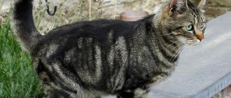
Cats... Many people's pets. Some people like red ones, some like black ones, some like mosaic ones. Others are attracted to Persians, Siamese cats or Egyptian cats. It's all a matter of taste.
However, the color of an animal, its exterior, character, diseases, pathologies, mutations depend not only on the breed or lifestyle, but also on the chromosome set (primarily on it), which is constant and definite.
And yet, how many chromosomes does a cat have, what are their numbers and functions? This will be discussed below.
Genome and chromosomes
It is extremely difficult to talk about how many chromosomes a cat has without basic knowledge of genetics.
A genome is a structure that contains the genetic information of an organism. Almost every cell contains a genome. But the chromosome contains all the information about the structure of the cell. A chromosome is a nucleoprotein structure in the nucleus of a eukaryotic cell. The chromosome contains a significant part of hereditary information, which is stored, implemented and transmitted to the future generation.
A chromosome is a structure in the cell nucleus made up of deoxyribonucleic acid (DNA) and proteins. It is worth recalling that DNA is a macromolecule that ensures storage, transmission from generation to generation, and the implementation of the genetic program for the development of a living organism.
There are two types of chromosomes in different organisms:
Eukaryotic type - characteristic of living organisms (eukaryotes), whose cells contain a nuclear membrane, DNA molecules in the nucleus and mitochondria.
Prokaryotic type - found in organisms whose cells lack a nuclear envelope, and DNA molecules are enclosed in histones (prokaryotes).
Externally, a chromosome looks like a long thread with stringed beads, each of which is a gene. In addition, the gene is located on its strictly fixed part of the chromosome - the locus.
What are chromosomes
XY Chromosomes
Chromosomes are structures found in the cells of living organisms. Each chromosome contains one twisted molecule of DNA - the main keeper of genetic data. Accordingly, chromosomes in living organisms are needed for:
- storage of hereditary information;
- transferring it through replication (doubling) of a DNA molecule and transferring its copy to daughter cells formed as a result of division.
A gene is a section of a DNA molecule that contains complete information about any trait or property of an organism.
Each species of animal or plant has a certain number of chromosomes, which does not change throughout life. They are arranged in pairs, so normally there is always an even number of them. An odd number means a chromosomal defect, for example, 47 chromosomes in a person (Down syndrome).
Number of chromosomes in animals
How many chromosomes are there in cat cells? Any living organism has homologous or paired chromosomes and haploid or unpaired (sex) chromosomes. The latter include the egg and sperm; they have a set of XX and XY, respectively. When dividing, they break up into X, X and X, Y. Depending on the new combination of the pair in the fertilized cell, the sex of the new organism (in our case, a kitten) will be determined.
To the question: “How many chromosomes are there in cat cells?”, genetics gives an exact answer. In a domestic cat, the chromosome set includes 19 pairs of chromosomes (18 paired and 1 unpaired: XX in females and XY in males). The total number of chromosomes in a cat is 38.
In other animals, the number of chromosomes is constant and individual for each species (for example, dogs have 78 chromosomes, horses have 64, cows have 60, and hare has 48). Recall that humans have 46 chromosomes.
Features of the karyotype of dogs and cats
Each cell in the body has a nucleus that stores genetic information. Its main part is contained in specific structures - chromosomes - connected chains of genes, which can be examined under a microscope at the stage of cell division.
The number and structure of chromosomes is a constant indicator specific to each type of living organism, which is called a karyotype. It determines the characteristics of inheritance of most of the characteristics and properties of an animal. A violation of their quantity or other changes can cause the development of hereditary diseases, the birth of non-viable individuals or, conversely, new species.
Each cell contains a constant paired number of identical chromosomes, characteristic of the species: a domestic cat has 38 (19 pairs), a dog has 78 (39 pairs). They determine the characteristics of the appearance, health and character of each individual. In germ cells there is only part (half) of this set, which is restored during fertilization.
All pairs of chromosomes, with the exception of one, have the same appearance (shape and size) and are responsible for the development of the same characteristics, while one pair contains chromosomes of different sizes, which are responsible for sexual characteristics:
- X - is large in size and determines the female gender,
- U – characterized by smaller size and denotes male gender.
The sex of the future offspring depends on the characteristics of their fusion: if during fertilization female and male cells with X chromosomes meet, a female individual develops, but if one of them contains a Y type, a male individual appears.
Karyotype and chromosome complex of a cat
A karyotype is a paired set of chromosomes with a specific number, size and shape for each animal species. The characteristics of each type of living organism are inherited by karyotype. For example, a karyotypic feature may be the presence of a trunk in elephants. The birth of a baby elephant without a trunk will be a deviation from the karyotypic norm, that is, a pathology.
All cells are paired, the future appearance, appearance, color, and character of the cat depend on them. The last - 19 pair contains sexual information and half of the chromosome set. During the process of fertilization, both parts join together to form a complete cell.
Principles of cat heredity
The information contained in chromosomes is the genotype. In turn, the external manifestations of certain characteristics are called phenotype.
Genes are arranged in pairs (one each from the mother and father) - alleles and are distinguished into the following subtypes:
- Dominant, that is, predominant. Thanks to this gene, certain characteristics of appearance are inherited by the offspring of the first generation.
- Recessive. Being a suppressed dominant, hidden for some time. Recessive genes combined with each other produce offspring that are unlike their parents.
Let's look at inherited traits:
- color;
- eye color;
- wool structure;
- size, shape and position of the ears;
- tail length and thickness;
- structure of the body and limbs.
Heredity of cats
There are 38 chromosomes in a cat's somatic cell, which contain DNA molecules with genotypic information. Genotypic manifestations that are reflected in the external appearance of a living organism are called phenotype. Phenotypic manifestations of kittens vary in color and size of the animal.
The offspring have paired genes - one gene from the female, and the other from the male. As you know, genes are divided into dominant (strong) and recessive (weak). Dominant genes are indicated in uppercase, Latin letters, recessive - in lowercase. Depending on their combination, homozygous (AA or aa) and heterozygous (Aa) types are distinguished. The dominant gene manifests itself in both homo- and heterozygous states. The recessive gene will manifest its characteristics only in the homozygous type (aa). This genetic knowledge is useful in calculating the traits of future kittens from the phenotypic expressions of their parents. Here it is important to know which gene responsible for the manifestation of a certain trait is recessive or dominant.
Animal color genes are located on the X chromosome, they are presented in the table below:
| a | Grey |
| b | Chocolate |
| c | Platinum, lilac |
| d | Ginger |
| e | Cream |
| f | Tortoiseshell |
| g | Tortoise blue cream |
| h | Turtle chocolate |
| j | Tortoiseshell lilac |
| n | Black |
| o | Sorel, honey |
| p | Yellow-brown |
| q | Tortoiseshell red-brown |
| r | Tortoiseshell tan |
| s | Smoky |
| w | White |
| y | Gold |
| x | Unregistered color |
Breeding wide-footed cats
Broad-footed Maine Coon breeds are bred in nurseries based on breeding programs, where the corresponding gene is passed on between generations. To produce offspring with polydactyly, one parent must have a foot anomaly - then the birth of offspring with polydactyly is possible with a probability of 56% (not all kittens in the litter are polydactylic).
If you breed two wide-legged animals, the likelihood of having polydact kittens increases by another 20%. Mating, where one parent is a homozygous polydact, leads to the fact that the entire litter will be polydactyl.
Polydactyly in cats
The polydactyly gene behaves unpredictably. It is expressed differently in offspring, regardless of the combination of fingers of the parents. In contrast to other dominant genes, it manifests itself in different ways in both homo- and heterozygous forms.
Breeders of broad-toed cats continue to conduct statistical work, trying to discover more accurate information about the likelihood of producing multi-toed kitten offspring.
Be sure to read:
Caring for an amazing raccoon cat - Maine Coon
Coat color
In a thousand genes of cats there are those that are responsible both for their coloring and for the mutation that leads to a change in the color and structure of the coat. A non-reproductive somatic cell contains in the proto-oncogene elements of a mutation in coat color, which inhibits the migration of melanoblasts. Therefore, the latter cannot penetrate the skin, and the pigment, accordingly, does not reach the hair of the coat. This explains the white coat of the animal.
Some melanoblasts manage to get into the hair follicles on the cat's head, and then colored spots appear on the fur. These cells may well reach the retina of the eyes: with a small number of melanoblasts, the eyes become blue, and with a large number, the animal’s pupils will be yellow.
The same chromosome is also responsible for coat color. The usual structural form of melanoblasts gives the animal a striped color. There are also semi-dominant changes, for example, in the Abyssinian tabi. Homozygous individuals do not have stripes, the color is uniform, and heterozygous individuals with this mutation are distinguished by stripes on the muzzle, paws, and tail. In the case of a recessive change, the transverse stripes on the animal’s fur are deformed into irregular lines on its back, appearing in powerful longitudinal stripes of black color.
A gene mutation affecting the enzyme tyrosinase leads to albinism. This occurs not only in cats, but also in other mammals.
Tyrosinase reduces its activity depending on the temperature of the cats - the lower it is, the more active the enzyme. In such cases, there is intense staining of the peripheral parts of the body: the nose, tips of the paws and tail, and ears in Burmese cats.
What heritable traits are transmitted by chromosomes?
Genetics is a baby in the scientific world. It is the same cosmos, only inside an individual. To date, scientists have been able to isolate a negligible number of genes responsible only for the simplest traits:
- wool (its color, structure and length);
- shape and location of the ears;
- eye color;
- body anatomy (tail, paws, height).
Diseases are also inherited. They are called genetic. Here are just a few of them:
- cardiomyopathy;
- Hypertrophic cardiomyopathy is a hereditary disease manifested in thickening of the walls of the ventricles of the heart, which leads to a decrease in their ability to contract porphyria - a disorder of hemoglobin synthesis;
- meningocele - brain herniation;
- von Willebrand disease - increased bleeding of the mucous membranes;
- retinal atrophy;
- polycystic kidney disease.
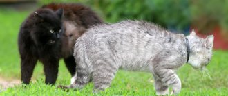
Mosaic of cats
The set of cat chromosomes responsible for coloring is localized on the X chromosome. Patterned cats are not uncommon, but there are still fewer three-colored cats than two-colored ones.
In this case, the color is determined by the alleles of the O gene:
O - affects the yellow (or red) color of the fur;
o - responsible for black color.
Tortoiseshell cats are heterozygous for this gene, their genotype is Oo.
Their yellow and black spots develop as a result of random inactivation in early embryogenesis of the X chromosome by the O or o allele. Cats can only be homozygous for this trait (OU - red or OU - black).
Tortoiseshell cats are extremely rare - they are characterized by the chromosomal constitution of XXXY and the genotype OoU. This is the reason for the rare birth rate of mosaic cats (or tortoiseshell cats).
Inheritance of the tricolor color of cats:
Color black – XB gene – genotype – XB XB; HVU;
Red color – Xb gene – genotype – Xb xb; HYU;
Tortoiseshell color – gene – XB; Xb – genotype XB; XH.
Mechanism of heredity
Some new breeds of animals have gained the right to life thanks to a gene mutation.
In each new generation, genetic mutations and redistribution of the chromosomes of both parents make it possible to reproduce more exotic and hardy cats. The biological codes of each organism contain a lot of information that scientists use for historical research. Sometimes this is the only way to find out the answers to questions about life many thousands of years ago.
Previously, historians believed that the animal was first domesticated in Egypt, but DNA analysis of buried cats proved this opinion wrong. The original genes of today's cats were inherited from the Wild African, which is their direct genetic ancestor. The domestic cat is a subspecies of the forest cat. She has the ability to hear even rustling sounds and sees perfectly in the dark. These and other natural data allow it to survive in any conditions by feeding on rodents or other small animals. Living for thousands of years with people, the cat still remains true only to itself.
The genes of cats already studied by geneticists are divided into 2 types:
- Strong (dominant). Indicated in capital letters. They are noticed in the first generations, since their activity does not depend on the presence in the body of other genes of the same trait.
- Weak (recessive). Marked with small signs. The development of a trait occurs only in the absence of other variations of such a gene and mainly manifests itself in the second and subsequent generations.
Each of the genes is responsible for different qualities in the body and some of them are interconnected. A single trait may be under the control of multiple alleles, making them difficult to fully study.
The size of the ears in animals is also determined by heredity.
Inherited traits in cats:
- parameters of the ears, as well as ear sizes;
- coat color and length;
- eye color;
- tail structure;
- presence of diseases.
White color of cats
White color at the chromosomal level is the absence of pigment. Pigment cells are blocked by one gene - W. If the genotype of cats contains recessive traits of this gene (ww), then the offspring will be colored, and if there is a dominant trait (WW, Ww) and at the same time in the genome of cats there will be many other designations of gene chromosomes (BOoSsddWw ), then we will still see a completely white cat. However, such cats can carry both spotting and patterning, but only if the offspring do not inherit the W gene.
Chromosomes of a cat with Down syndrome
This disease occurs not only in humans, but also in animals, cats are no exception.
There are many stories and photographs from the lives of such animals on the Internet. Just like people, such animals can live and be active, but visually they differ from healthy ones. Like people, such animals need certain care, care and treatment.
To the question: “How many chromosomes does a Down cat have?”, one can definitely answer: 39.
Down syndrome occurs when another extra chromosome appears in the gene set of chromosome molecules - an odd one. In the case of cats, this is chromosome 39.
A cat with an extra chromosome is rare in nature for the simple reason that the animal does not use drugs, alcohol, or smoke, i.e. the provoking causes of gene mutation are excluded. But still, this is a living organism, sometimes it also has malfunctions.
Scientists and biologists do not have a definite opinion about the extra chromosome. Some say that this cannot happen, others say that it can, and still others claim that this occurs when an animal is artificially bred as an experimental animal.
A cat with 20 chromosomes (the twentieth pair of chromosomes is an extra one) can be found, but it has virtually no chance of reproducing healthy offspring. This, of course, does not mean that such an animal cannot be loved. They are quite cute, but a little unusual, different, but still they are alive. For example, a cat with this syndrome (Maya from America) became the favorite of her owners (Harrison and Lauren). They created their own page on Instagram for the cat and regularly post photos and videos of it. Maya has become a favorite of Internet users; she is quite active and cheerful, although she suffers from shortness of breath and constantly sneezes. But no one bothers her to live for her own pleasure and for the pleasure of her masters.
By the way, you should not confuse a cat's Down syndrome with genetic mutations that lead to physical changes (deformations) in the animal's faces. This occurs in nature more often than Down's disease, and is caused by crossing between cats-relatives (interbreeding). If there are many animals of the same kind in the offspring, then sooner or later physiological changes will occur not only in the appearance of the animals, but will also affect their development as a whole. While breeders can control this, the owners of cats running around their yards are practically unable to track it. Some people abort such offspring, others, on the contrary, take this philosophically and also love their pets.
Hereditary factors
The karyotype plays a determining role in the heredity of cats. The somatic cell of an animal contains 38 chromosomes, which, in turn, consist of smaller particles - DNA genes. These genes carry information that will certainly be passed on to subsequent generations, with the exception of cases where we are talking about a castrated individual. Any external manifestations in a kitten that are characteristic of its parents are called a phenotype.
Since the number of chromosomes (with the exception of the sex chromosome) is paired, this means that information is transmitted from both the mother and the father. However, the amount of information inherited may vary. It depends on which gene expresses itself to a greater extent.
It is known that they can be dominant and recessive. There is a significant difference here:
| Dominant | Recessive |
| It is the leader, and accordingly, the information stored in it appears in the first generation of the inheritance. Indicated in capital Latin letters | It is not as strong as the dominant one, and therefore, as a rule, manifests itself only in subsequent generations, for example, in the second or even third. Indicated in lowercase Latin letters. |
Depending on the combination of genes, homozygous and heterozygous types are determined. The first is AA or aa, that is, a combination of two dominant or two recessive genes. The second is Aa, a combination of dominant and recessive genes. The stronger gene actively manifests itself in any variant of the combination, but the weaker one - only in homozygous aa.
It is noteworthy that if cats give birth to a kitten with the homozygous type aa, then it may be completely different from its parents, since weak genetics will manifest itself only in its descendants. That is, for example, his “grandchildren” will look like the cat’s parents.
It is the chromosomes that transmit by inheritance such important information as:
- coat color and type;
- eye color;
- tail length and thickness;
- size and location of the ears, shape of the auricle;
- predisposition to certain diseases.
Coat color
Among the set of genes in cats, there are those that are directly responsible for the color of the coat and its structure. This scheme works like this: a non-reproductive somatic cell contains a certain number of such genes. Their task is to inhibit the spread of melanoblasts. This is a pigment that, in fact, gives the coat a particular color. If the gene prevents melanoblasts from reaching the hair, then the hair in this place does not color and remains white. If this gene is not present in certain cells, it means that migration of melanoblasts is possible here, as a result of which the coat color occurs. This is how characteristic spots appear on the animal’s body.
Among the set of genes in cats there are those that are directly responsible for coat color and its structure.
The normal pigment, which is not influenced by genetic information, gives the striped color. But genes can cause melanoblasts to color their fur in different shades. It all depends on the karyotype. Heterozygous cats will have a striped color, while homozygous cats will have a uniform color.
Sometimes a gene mutation is possible, as a result of which the production of the tyrosinase enzyme decreases or even stops. As a result, the animal develops albinism.
Mosaic
When a kitten has one or two colors, this is a normal phenomenon, familiar to everyone. But sometimes a pet can look like a real mosaic, because its fur combines three or even more shades. Why is this happening?
Naturally, it’s all about the genes that make up these structures. The genes responsible for coloring are contained on the X chromosome. The reason for the mosaic pattern is that the chromosome consists of various genes that “color” the coat. That is, there is nothing extraordinary here, although it is a comparative rarity.
Sometimes a pet can look like a real mosaic, because its fur combines three or even more shades
How many lives does a cat have?
Everyone knows that in 1996 the first cloning was carried out in the world (the famous Dolly the sheep). Five years later, scientists cloned a cat and gave it a name – Kopirka (in Russian) or Carbon Copy (in Latin).
For cloning, a gray-red tortoiseshell cat, Rainbow, was taken. Eggs and somatic cells were extracted from Rainbow's ovaries. Nuclei were removed from all eggs and replaced with nuclei isolated from somatic cells. Electroshock stimulation was then performed and the reconstructed eggs were transplanted into the uterus of a gray tabby cat. It was this surrogate mother who gave birth to Copier.
But Kopirka had no red spots. The study revealed the following: in the genome of a cat (female) there are two X chromosomes, which are responsible specifically for the color of the animal.
In a fertilized cell (zygote), both X chromosomes are active. During the process of cell division and further differentiation in all cells of the body, including future pigment cells, one of the X chromosomes is inactivated (that is, the cell’s activity is lost or greatly reduced). If a cat is heterozygous (for example, Oo) for the color gene, then in some cells the chromosome carrying the red color allele can be inactivated, in others - the chromosome carrying the black color allele. Daughter cells strictly inherit the state of the X chromosome. As a result of this process, the tortoiseshell coloration is formed.
When a cat was cloned in the nucleus of a reconstructed egg extracted from an ordinary somatic cell of a calico cat, complete reactivation (restoration of viability or activity) of the switched off X chromosome did not occur.
Complete reprogramming of the chromosome nucleus does not occur when cloning a living organism (in this case, a cat). It is likely that this is why cloned animals get sick and cannot always produce healthy offspring. The carbon copy is still alive. She became the mother of three adorable kittens.
Human sex cells, chromosomes, fertilization
Sex cells - gametes (from the Greek gametes - “spouse”) can be found already in a two-week human embryo. They are called primordial germ cells . At this time, they are not at all similar to sperm or eggs and look exactly the same. It is not possible to detect any differences inherent in mature gametes at this stage of embryo development in primary germ cells. This is not their only feature. Firstly, primary germ cells appear in the embryo much earlier than the sex gland itself (gonad), and secondly, they arise at a considerable distance from the place where these glands will form later. At a certain moment, an absolutely amazing process occurs - the primary germ cells rush together to the gonad and populate, “colonize” it.
After the future gametes enter the gonads, they begin to divide intensively, and their number increases. At this stage, germ cells still contain the same number of chromosomes as “bodily” ( somatic ) cells - 46. However, to successfully carry out their mission, germ cells must have 2 times fewer chromosomes. Otherwise, after fertilization, that is, the fusion of gametes, the cells of the embryo will contain not 46, as established by nature, but 92 chromosomes. It is not difficult to guess that in subsequent generations their number would progressively increase. To avoid this situation, the developing germ cells undergo a special division, which in embryology is called meiosis (Greek meiosis - “reduction”). As a result of this amazing process the diploid (from the Greek diploos - “double”) set of chromosomes is, as it were, “pulled apart” into its constituent single, haploid sets (from the Greek haploos - single). As a result, from a diploid cell with 46 chromosomes, 2 haploid cells with 23 chromosomes are obtained. Following this, the final stage of the formation of mature germ cells begins. Now, in a haploid cell, the existing 23 chromosomes are copied and these copies are used to form a new cell. Thus, as a result of the two divisions described, 4 new ones are formed from one primary germ cell.
Moreover, in spermatogenesis (Greek genesis - origin, development), as a result of meiosis, 4 mature sperm with a haploid set of chromosomes appear, and in the process of egg formation - in oogenesis (from Greek oon - “egg”) only one. This happens because the egg cell does not use the second haploid set of chromosomes formed as a result of meiosis to form a new mature germ cell - an oocyte, but “throws” them out as “extra” in a kind of “garbage container”, which is called a polar body. The first division of the chromosome set is completed in oogenesis with the release of the first polar body just before ovulation. The second replication division occurs only after the sperm penetrates the egg and is accompanied by the release of the second polar body. For embryologists, polar bodies are very important diagnostic indicators. The first polar body is present, which means the egg is mature, the second polar body appears, and fertilization has occurred.
Primary germ cells found in the male gonad do not divide for the time being. Their division begins only during puberty and leads to the formation of a cohort of so-called diploid stem cells, from which sperm are formed. The supply of stem cells in the testicles is constantly replenished. Here it is appropriate to recall the feature of spermatogenesis described above - 4 mature sperm are formed from one cell. Thus, after puberty, a man produces hundreds of billions of new sperm throughout his life.
The formation of eggs proceeds differently. Having barely populated the gonad, the primary germ cells begin to divide intensively. By the 5th month of intrauterine development, their number reaches 6-7 million, but then mass death of these cells occurs. In the ovaries of a newborn girl there are no more than 1-2 million of them, by the age of 7 - only about 300 thousand, and during puberty 30-50 thousand. The total number of eggs that will reach a mature state during puberty will be even less. It is well known that during one menstrual cycle, only one follicle usually matures in the ovary. It is easy to calculate that during the reproductive period, which lasts for women 30-35 years old, about 400 mature eggs are formed.
If meiosis in spermatogenesis begins during puberty and is repeated billions of times during a man’s life, in oogenesis the forming female gametes enter meiosis during the period of intrauterine development. Moreover, this process begins almost simultaneously in all future eggs. It begins, but does not end! Future eggs reach only the middle of the first phase of meiosis, and then the division process is blocked for 12 - 50 years! Only with the advent of puberty will meiosis continue in oogenesis, and not for all cells at once, but only for 1-2 eggs monthly. The process of meiotic division of the egg will be completed, as mentioned above, only after its fertilization! Thus, the sperm penetrates into an egg that has not yet completed division and has a diploid set of chromosomes!
Spermatogenesis and oogenesis are very complex and largely mysterious processes. At the same time, their subordination to the laws of interrelation and conditionality of natural phenomena is obvious. To fertilize one egg in vivo (lat. in a living organism), tens of millions of sperm are needed. The male body produces them in gigantic quantities almost throughout his life.
Carrying and giving birth to a child is an extremely difficult burden on the body. Doctors say that pregnancy is a health test. How a child will be born directly depends on the state of health of the mother. Health, as you know, does not last forever. Old age and illness, unfortunately, are inevitable. Nature gives a woman a strictly limited, irreplaceable number of germ cells. A decrease in fertility develops slowly, but gradually along an incline. We receive clear evidence that this is indeed the case by daily assessing the results of ovarian stimulation in ART programs. Most of the eggs are usually used up by age 40, and by age 50 the entire supply is completely depleted. Often, so-called ovarian depletion occurs much earlier. It should also be said that the egg is subject to “aging”; over the years, its ability to fertilize decreases, and the process of chromosome division is increasingly disrupted. Having children at a late reproductive age is risky due to the increasing risk of having a child with a chromosomal abnormality. A typical example is Down syndrome, which occurs due to an extra 21 chromosome remaining during division. Thus, by limiting the reproductive period, nature protects the woman and takes care of healthy offspring.
According to what laws does chromosome division occur? How is hereditary information transmitted? In order to understand this issue, we can give a simple analogy with cards. Let's imagine a young married couple. Let's call them conventionally - He and She. Each of its somatic cells contains chromosomes of the black suit - clubs and spades. He received a set of clubs from six to ace from his mother. A set of spades - from my dad. In each of its somatic cells, red chromosomes are diamonds and hearts. She received a set of diamonds from six to ace from her mother. A set of worms - from my dad.
In order to obtain a sex cell from a diploid somatic cell, the number of chromosomes must be halved. In this case, the sex cell must contain a complete single (haploid) set of chromosomes. Not a single one should get lost! In the case of cards, such a set can be obtained as follows. Take one at random from each pair of black cards and thus form two single sets. Each set will include all cards of the black suit from six to ace, however, what kind of cards these will be (clubs or spades) is determined by chance. For example, in one such set the six may be a spades, and in another it may be a club. It’s not hard to imagine that in the example with cards, with such a choice of a single set from a double set, we can get 2 combinations to the ninth power - more than 500 options!
In the same way, we will make a single set of her red cards. We will get more than 500 different options. From his single and her single set of cards we will make a double set. It will turn out to be, to put it mildly, “variegated”: in each pair of cards, one will be of a red suit, and the other will be black. The total number of such possible sets is 500×500, that is, 250 thousand options.
Nature does approximately the same thing, according to the law of random sampling, with chromosomes during the process of meiosis. As a result, from cells with a double, diploid set of chromosomes, cells are obtained, each of which contains a single, haploid complete set of chromosomes. Let's say that as a result of meiosis, a sex cell is formed in your body. Sperm or egg - in this case it does not matter. It will definitely contain a haploid set of chromosomes - exactly 23 pieces. What exactly are these chromosomes? Let's take chromosome 7 as an example. This could be the chromosome you received from your father. It could just as likely be a chromosome you got from your mother. The same is true for chromosome No. 8, and for any other.
Since in humans the number of haploid chromosomes is 23, then the number of possible variants of sexual haploid cells formed from diploid somatic cells is equal to 2 to the power of 23. This results in more than 8 million variants! During the process of fertilization, two germ cells unite with each other. Therefore, the total number of such combinations will be 8 million x 8 million = 64,000 billion options! At the level of a pair of homologous chromosomes, the basis of this diversity looks like this. Let's take any pair of homologous chromosomes of your diploid set. You received one of these chromosomes from your mother, but it could be from either your grandmother or your maternal grandfather. You received the second homologous chromosome from your father. However, it can again be, regardless of the first chromosome, either your grandmother’s or your paternal grandfather’s. And you have 23 pairs of such homologous chromosomes! This results in an incredible number of possible combinations. It is not surprising that one pair of parents gives birth to children who differ from each other in both appearance and character.
By the way, a simple but important conclusion follows from the above calculations. Every person currently living, or who has ever lived in the past on Earth, is absolutely unique. The chances of a second one appearing are almost zero. Therefore, there is no need to compare yourself with anyone. Each of you is unique, and that makes you interesting!
However, let's return to our reproductive cells. Each diploid human cell contains 23 pairs of chromosomes. Chromosomes from 1 to 22 pairs are called somatic and they are the same in shape. The chromosomes of the 23rd pair (sex chromosomes) are the same only in women. They are designated by the Latin letters XX. In men, the chromosomes of this pair are different and are designated XY. In the haploid set of an egg, the sex chromosome is always only X, while the sperm can carry either an X or a Y chromosome. If the egg is fertilized by X sperm, a girl will be born, if Y sperm, a boy will be born. It's simple!
Why does meiosis in an egg take so long? How is a monthly selection of a cohort of follicles that begin their development and how is the leading, dominant, ovulatory follicle selected from them, in which the egg will mature? Biologists do not yet have clear answers to all these difficult questions. The process of formation of mature eggs in humans awaits new researchers!
The formation and maturation of sperm, as already mentioned, occurs in the seminiferous tubules of the male reproductive gland - the testicles . The formed sperm has a length of about 50-60 microns. The sperm nucleus is located in its head. It contains the paternal hereditary material. Behind the head there is a neck, in which there is a large convoluted mitochondrion - an organelle that ensures the movements of the tail. In other words, this is a kind of “energy station”. There is a “cap” on the head of the sperm. Thanks to it, the shape of the head is oval. But, it’s not about the form, but about what is contained under the “cap”. This “cap” is actually a container and is called an acrosome , and it contains enzymes that are capable of dissolving the shell of the egg, which is necessary for the sperm to penetrate inside - into the cytoplasm of the egg. If the sperm does not have an acrosome, its head is not oval, but round. This sperm pathology is called globulospermia (round-headed sperm). But, again, the trouble is not in shape, but in the fact that such a sperm cannot fertilize an egg, and a man with such a disorder of spermatogenesis was doomed to childlessness until the early 90s of the last century. Today, thanks to ART, infertility in these men can be overcome, but we will talk about this later in the chapter on micromanipulation, in particular ICSI.
The movement of the sperm is carried out due to the movement of its tail. The speed of sperm movement does not exceed 2-3 mm per minute. It would seem not much, however, in 2-3 hours in the female reproductive tract, sperm travel a distance 80,000 times greater than their own size! If a person were in the place of the sperm in this situation, he would have to move forward at a speed of 60-70 km/h - that is, at the speed of a car!
The sperm in the testicle are immobile. They acquire the ability to move only by passing through the vas deferens under the influence of the fluids of the vas deferens and seminal vesicles, and the secretion of the prostate gland. In the female genital tract, sperm remain motile for 3-4 days, but they must fertilize the egg within 24 hours. The entire development process from a stem cell to a mature sperm lasts approximately 72 days. However, since spermatogenesis occurs continuously and a huge number of cells enter it at once, the testicles always contain a large number of sperm at different stages of spermatogenesis, and the supply of mature sperm is constantly replenished. The activity of spermatogenesis varies from person to person, but decreases with age.
As we have already said, eggs are located in the follicles of the ovary. As a result of ovulation, the egg enters the abdominal cavity, from where it is “caught” by the fimbriae of the fallopian tube and transferred to the lumen of its ampullary section. This is where the egg meets the sperm.
What structure does a mature egg have? It is quite large and reaches 0.11-0.14 mm in diameter. Immediately after ovulation, the egg is surrounded by a cluster of small cells and a gelatinous mass (the so-called corona radiata ). Apparently, in this form it is more convenient for the fimbriae of the fallopian tube to capture the egg. In the lumen of the fallopian tube, with the help of enzymes and mechanical action (beating of the cilia of the epithelium), the egg is “cleaned” from the corona radiata. The final release of the egg from the corona radiata occurs after it meets the sperm, which literally stick around the egg. Each sperm secretes an enzyme from the acrosome that dissolves not only the corona radiata, but also acts on the membrane of the egg itself. This shell is called the pellucida, which is what it looks like under a microscope. By secreting the enzyme, all sperm strive to fertilize the egg, but the zona pellucida will allow only one of them to pass through. It turns out that by rushing towards the egg and acting on it collectively, the sperm “clear the way” for only one lucky person. The role of the zona pellucida is not limited to the selection of sperm; in the early stages of embryo development, it maintains the ordered arrangement of its cells (blastomeres). At some point, the zona pellucida becomes tight, it ruptures and hatching (from the English hatching - “hatching”) - the hatching of the embryo. The embryo is ready for implantation into the endometrium.