Many types of yeast fungi are opportunistic microflora that constantly live on the skin and mucous membranes of the animal’s body. They do not cause pathology and live in symbiosis with the host until a sharp decrease in immunity occurs. During this process, microorganisms begin to multiply intensively, which leads to the occurrence of diseases. A striking example is the development of Malassezia in cats.
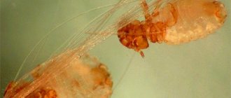
Malassezia globosa fungus under a microscope
Malassezia dermatitis is caused by the yeast Malassezia pachydermatis. For the most part, they inhabit the external auditory canal, interdigital folds, anal glands in males, vagina in females, and the rectum. Only weakened animals suffer from this disease; with a timely diagnosis, treatment initiated and proper care, recovery occurs in a short period of time.
What it is?
Malassezia pachydermatis is a type of yeast. In the vast majority of cases, they are part of the normal microflora of the skin and can be found in any animal. But sometimes something happens, after which harmless yeast suddenly becomes active and causes a lot of problems. Typically, these fungi are found in the external auditory canal; they are found in the anal sinuses, vagina and rectum. Malassezia can affect animals of all breeds and ages; gender differences also do not play any role.
Why can this microflora become active? In general, the reasons in this case are similar for all fungal infections. Any chronic and, in particular, hereditary autoimmune diseases that lead to a prolonged decrease in immune status are classic predisposing factors. Cats suffering from any bacterial infection, allergy or seborrhea almost always have inflamed and irritated skin. Perhaps a better “springboard” for mushrooms cannot be found.
It should be noted, however, that this kind of infection in cats is very rare, but the most common of them is Malassezia in the ears of a cat. This is due to the fact that the parasite feels most comfortable inside the ear canal.
Causes of the disease
Malaceziosis is, as a rule, a secondary disease that appears against a background of weakened immunity. The reasons for this may be the following:
• infectious disease;
• food allergies;
• external and internal parasites;
• hormonal imbalance;
• diseases of the digestive or urinary systems;
• mechanical damage to the skin (bites, scratches, wounds).
The Malassezia fungus is considered opportunistic. It is found in small quantities on the skin of all cats and does not cause disease under normal conditions. When the protective barriers are weakened, caused by allergies, injuries, infections or other reasons, it is activated and begins to multiply intensively. As a result, the lipid-carbohydrate balance of the skin is disrupted and a disease called malasseziosis occurs.
Therapy
Treatment of Malassezia in cats can be carried out in several ways. But let us warn you once again that before starting therapy it is very important not only to identify the pathogen, but also to make sure that it is the one that caused the disease. If treatment over a long period of time does not produce a positive effect at all, a full examination of the animal should be carried out in order to identify the true root cause of the pathology.
To create an unsuitable environment for the fungus to live, it is necessary to remove excess fat from the cat's skin. For this, both specially developed shampoos and more conventional products can be used. A 1% solution of chlorhexidine has proven itself well. In severe cases, the concentration may be increased. Products containing benzoyl peroxide and sulfur are also used.
Diagnostics
The diagnosis of malasseziasis is made based on the following examination results of the animal:
• medical history data – the patient’s living conditions and illnesses he has suffered;
• identifying characteristic symptoms of the lesion;
• detection of a large number of Malassezia cells in skin scrapings under a microscope and colonies of the fungus during culture.
Most often, malacesia occurs in weakened cats or in kittens during the transition from breastfeeding to solid food .
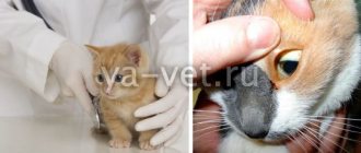
During the examination, if characteristic symptoms of the disease are detected, scrapings are taken from the affected areas of the cat’s skin, which are examined under a microscope. At the same time, inoculations are made on the nutrient media.
Under a microscope, Malassezia looks like a groundnut. The cells are arranged in groups of several, attached to a nutrient substrate - the epidermis or hairs of wool. The diagnosis of malassezia is made if more than 10 microorganisms are detected in 15 points of the test sample at 1000x magnification. When sown on Sabouraud agar, Malassezia forms convex, well-defined, spherical colonies of yellow or yellowish-brown color.
Since malacesia is a secondary disease, in parallel with cytological studies, tests are carried out for the presence of allergies, and blood is taken for biochemistry. If there are no results, a comprehensive examination of the animal is carried out to identify the main cause of the disease.
Malassezia in animals: what are the advantages of veterinary
Malassezia should be treated according to the recommendations of a specialist - a veterinarian. Before turning to any veterinary center for help, it is recommended to soberly evaluate its advantages and decide for yourself whether it is good enough for you. Veterinary, has the following advantages:
- Modern equipment, both in the veterinarian’s office and in the laboratory;
- Qualified specialists who regularly update their knowledge with new knowledge;
- Positive attitude towards animals;
- Possibility of calling a doctor at home;
- Possibility of testing at home;
All these advantages set us apart from other veterinary centers and allow us to fully experience all the delights of a modern approach to the provision of veterinary services.
Treatment
First of all, when starting treatment, it is necessary to eliminate the provoking factor:
- abolition of antibacterial therapy and normalization of nutrition for candidiasis,
- treatment of bacterial infections in mold fungal mycosis,
- elimination of excessive humidity, atopy in Malassezia.
Nutrition should be complete, with sufficient content of all nutrients. Additionally, vitamin preparations and a course of immunostimulants (Gamavit) are administered.
For all skin diseases, it is recommended to administer a solution of potassium iodide.
Candidiasis
Therapy for candidiasis should be comprehensive and include both systemic drugs and local therapy.
For application to the skin, use Wilkinson's ointment, zoomukol, nizoral ointment.
For systemic therapy, use nizoral 1/3 tablet per day for a week or levorin.
Mycosis by mold fungus
Treatment must be long-term, lasting at least 6 weeks. During it, exoderil is used for external use to lubricate the lesions, and nizoral or amphotericin. When fistulas form on nodules, the formations are cut out.
Malassezia
Just like other fungal infections, it is treated with special antifungal shampoos or solutions based on imidazole, miconazole or chlorhexidine for more than 2 weeks. For more thorough treatment, it is recommended to trim the animal. Especially long-haired breeds.
Ketoconazole is given orally with the obligatory parallel use of hepatoprotectors (covertal).
To relieve itching, antihistamines - suprastin - are added to therapy. To prevent infection from scratching, special collars and blankets are used.
Antibiotics are used when a primary source of bacterial infection is attached or present.
Diagnosis
Malacia dermatitis can be suspected if the skin disease is accompanied by itching, redness, inflammation and an unpleasant odor and localization. Clinical signs can complicate and vary keratinization defects and allergic skin diseases.
It has been proven that cats of allergenic breeds suffer from Malacia dermatitis twice as often.
When making a diagnosis for dermatitis caused by Malacesia, special attention is paid to persistent clinical manifestations and, first of all, to an increase in the population of M. pachidermatis on the damaged skin and a positive response to adequate antifungal therapy.
Typically, fungi are identified by cytological methods, although biopsy and culture may be more effective in providing information.
Yeast is isolated from the skin, as a rule, by taking smears, although the quantitative method is more informative. The contact method is most suitable for clinical practice. To do this, small agar plates should be pressed onto the affected skin for about ten seconds and then incubated for 3.7 days at 32 to 37 degrees Celsius to count the colonies. You can also use Dixon's dextrose agar, which supports the growth of more lipophilic variants of M. pachidermatis and makes it possible to isolate Malacesia species found in cats.
Malacia dermatitis is diagnosed by contact method
In most cases, if the cat is healthy, the amount of yeast in the groin and armpits is usually less than one colony-forming unit per 1 square cm. however, in the interdigital spaces and lip folds their number may be higher.
In addition, “breed” differences in population size can normally be observed. The population density in the armpits of some healthy representatives of some breeds exceeds the above-mentioned norm by ten times! This emphasizes how important response to treatment is as a diagnostic criterion, which is the most evidence-based.
Symptoms
Fungal infections can have different localizations, depending on the type of mycosis. It is most often located on the skin of the body, muzzle, in the ears and between the fingers. Manifestations of the infection itself range from scaly rashes to weeping ulcers and erosions.
Candidiasis
It has several basic forms:
- Intestinal is the most common type of illness. It manifests itself with characteristic symptoms of gastroenteritis - diarrhea, vomiting. Rashes may also appear on the mucous membranes of the mouth and conjunctiva. Small ulcers, redness and swelling of the tissues are found there. The mucous membrane is covered with a dense white coating.
- Pulmonary is the rarest form. Mainly accompanies bacterial infections with severely weakened immunity. It is characterized by pneumonia with copious sputum, shortness of breath and heavy shallow breathing due to chest pain.
- Skin - manifests itself in the form of rounded bald patches. The lesions are located on the legs and back. The skin inside the alopecia has a dirty gray tint and itches. Because of the latter fact, a bacterial complication often occurs when scratching, suppuration and ulceration appear.
- Vaginal - white, flaky discharge appears from the genital tract. When examining the vagina, the mucous membranes are covered with a white coating.
Manifestations for all forms of the disease are often vague; a combination of several forms at the same time is possible, especially in immunodeficiency.
Mold mycosis
Depending on the entrance gate of infection, there are 2 types:
- Pulmonary - enters the body by inhaling the fungus; usually the disease occurs as a complication of a bacterial infection. Nodular formations are observed in the lungs, hemorrhages are possible. The disease is accompanied by sneezing, apathy and anorexia, as well as fever and cough. A distinctive feature is parallel damage to the conjunctiva and nasal mucosa in the form of rhinitis.
- Skin - first, small nodules, about 3 centimeters in diameter, form at the site of abrasions. On top they are covered with dark, dead epithelium. After a few days, the formations are opened with the formation of fistulas and ulcers.
An intestinal form is also possible, which occurs with colitis, ulcerative-necrotic lesions of the gastrointestinal mucosa.
Malassezia
It can be localized and generalized. Typically located in the outer ear, between the fingers, on the face and abdomen. The affected skin is bright brown with a red tint and severe itching. Because of this, when scratching, a bacterial infection occurs, leading to the formation of ulcers, erosions, plaques and erythema. The appearance of an unpleasant sour odor from a cat is typical.
The main food for fungus is sebum, so you can often find it on the chin. Its presence there causes cat acne, the fur becomes dull and hangs in icicles. Over a long period of time, the skin becomes rougher and areas of alopecia appear.
Prevention
- Rational, balanced nutrition with high-quality food. It is best if your veterinarian selects the menu for you.
- Course prophylactic use of multivitamin preparations.
- Avoid frequent bathing and ear cleaning. Optimally for a cat - once every three months.
- Limiting contact with stray animals and going outside.
- Use only individual grooming items.
- Wash the bedding regularly.
- Vaccination.
- It is not advisable to keep your pet in a room with high humidity.
Clinical signs
So far, it has not been clarified whether there is a gender difference in the susceptibility to diseases caused by Malacesia.
Clinical description of dermatitis caused by Malacesia:
- First of all, it is seborrhea, peeling, hair loss, redness;
- Local or extensive skin lesions;
- Itching of varying degrees, up to the most severe;
- If the disease is chronic, lichenization and hypermentization often develop.
The most “popular” places are the interdigital space, abdomen, armpits, lower neck, mouth area and external auditory canal.
Often, clinical signs are observed together with allergic skin lesions, especially with atopy.
Itching is a sign of dermatitis