The author of the article is Kira Vadimovna Chernaya, a veterinary therapist at the IVC MBA.
Calcium oxalate (OxCa) is a salt of the alkaline earth metal calcium and the organic dibasic oxalic acid. Stones of this type are quite sharp, polygonal, due to which they strongly scratch the wall of the bladder, renal pelvis, ureters and urethra, causing pain and causing great damage to the mucous membrane. The detection of crystals of these stones in a urine test indicates a disturbance in the metabolism of oxalic acid salts and severe acidification of the urine. The formation of calcium oxalates often occurs in stressful situations, when the body does not have enough fluid for normal urinary function of the kidneys, with a weakly acidic urine reaction.
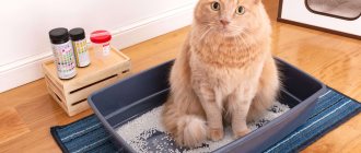
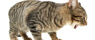
Cats with urolithiasis (presence of stones in the bladder) experience classic symptoms of feline cystitis: strained urination (sometimes even with vocalization due to pain), blood in the urine, diuresis (urination) in inappropriate places, licking of the genitals, frequent sitting down. Very often, cats pee on their owners' beds because they consider this place to be the safest in the world and try to help themselves in this way. In severe cases, usually in males, acute urinary retention occurs. Symptoms may be complex or absent altogether until a certain point. Usually, owners go to the veterinary clinic when the cat often sits on the tray for a long time, trying to pee, but 2-3 drops come out, sometimes with blood. The animal may scream, and sometimes there are puddles in different parts of the apartment.
Cats with nephrolithiasis (the presence of stones in the renal pelvis) and ureterolithiasis (the presence of stones in the ureters) may have no clinical signs of the disease at all, since cats rarely show chronic pain. Sometimes owners come to the clinic with complaints of apathy, lethargy, pain in the lumbar region of their pet, sometimes the cat may refuse to eat and vocalize.
How to find these stones?
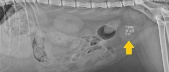
To detect calcium oxalates, radiography and ultrasound diagnostics are performed. Because of the large amounts of calcium in the joints of these stones, they “glow” on x-rays, almost like bone structure.
There may be no symptoms at all; often these stones are an accidental discovery, for example, during an X-ray examination of the abdominal organs to identify other pathologies, or during an ultrasound examination at a medical examination. But other types of uroliths (stones) can also be radiopaque, for example, struvites, cystines, calcium phosphates, magnesium-ammonium phosphates and others. Having identified stones in the bladder, we cannot be one hundred percent sure what we are faced with. For verification, it is recommended to surgically remove all found stones (one or more) and examine them in the laboratory.
The operation can be laparotomic (through an incision in the abdominal wall) or endoscopic (cystoscopy), but the second option is not suitable for everyone, due to the characteristics of the urinary system in cats - the urethra is thin and long, which does not allow such a procedure to be carried out in males. Veterinary surgery is undoubtedly evolving; your surgeon will choose the best option and communicate it to you. Unfortunately, there is no dietary food that will help dissolve stones of this type, there is only a maintenance diet from various manufacturers, but currently active development of food suitable for cats with calcium oxalate-type urolithiasis is underway.
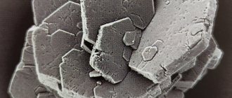
Calcium oxalate stones removed surgically
Dissolution of struvite stones
Drugs are used to reduce the urine acidity level to less than 6.0, but more often individually selected dietary diets are used that promote the dissolution of struvite. In dogs consuming these diets, the amount of protein, phosphate and magnesium entering the body is reduced and the amount of sodium increases. As a result of consumption of this diet, the volume of urine excreted increases, the urine becomes unsaturated, and this is an unfavorable environment for crystallization. When taking specialized diets, no other food or treats are recommended, and access to drinking water should be available around the clock.
Why do these stones appear? What else do we know about them?
- Twenty-five years ago, calcium oxalates were not found in cats, most often stones of the struvite type (a matrix of ammonium-magnesiophosphates) were detected.
- The veterinary food industry produced food to maintain the urological health of cats, acidifying the products to prevent the occurrence of struvite urolithiasis. In general, it worked - the number of cats with diseases of the lower urinary system decreased significantly. The trade-off was that under the influence of an acidic environment, calcium oxalates began to form, since at this pH (acid-base environment) calcium loss increased. Currently, the majority of kidney and ureteral stones in cats are calcium oxalates.
- Burmese and Himalayan cats are genetically predisposed to this type of urolithiasis.
- Most often, this disease occurs between the ages of 5 and 14 years.
- 35% of cats with calcium oxalate bladder stones have elevated calcium levels in the blood (hypercalcemia). It is advisable to examine the level of ionized calcium in the blood rather than the total level, as the result will be more accurate and indicative.
- In urine analysis, crystals of struvite (triple phosphates) are more often detected than oxalates, this is due to the strong structure of the latter.
- Cats with this pathology rarely have concomitant infections of the urinary system, and most often the urine density is high, that is, the urine is poorly diluted.
- Each animal has its own individual reaction to various substances, and even to suture material. If a surgical operation was performed to remove bladder stones, tumors, etc., and for some reason inflammation occurred along the suture, accumulations of calcium oxalate crystals may begin to form at this site, leading, in the future, to oxalate-type urolithiasis. Unfortunately, it is impossible to predict such a complication one hundred percent, which is why it is recommended to periodically conduct ultrasound, x-rays and monitor a general urine test for the occurrence of such problems.
- Urolithiasis of the oxalate type can occur due to metabolic acidosis (due to other problems, for example, diabetes, shock, trauma, ethylene glycol poisoning), systemically leading to active oxidative processes.
Is stone removal only surgical?
- Most often, such patients undergo cystotomy (abdominal surgery with an incision in the bladder) to remove stones, which are subsequently sent for research. This is the fastest and easiest method, used by most surgeons.
- Cystoscopy is considered a less invasive method. Flexible thin endoscopic instruments (cystoscopes) are used. This method of stone removal is indicated only for small stones, and is performed only on cats (females). Laser lithotripsy is sometimes used to break up large stones, in which case, if the procedure is successful, the stones may turn into sand and pass on their own, or they can also be removed endoscopically. In the Russian Federation, laser lithotripsy is performed in a few clinics and is not considered necessary due to the availability of simpler methods and the high cost of this procedure.
- If the diameter of the stones is small enough that they are actually sand, you can try bladder lavage under sedation. The patient is given anesthesia, a urethral catheter is installed, and the bladder is washed with Sodium Chloride. You can place the patient vertically, head up, then under the force of gravity and pressure from the solution, the stones can pass through the urethra through the catheter. This procedure is carried out quite rarely, as it has rather low efficiency.
- In the presence of massive stones, a diet for dissolving sand in the bladder is not indicated; it simply does not work.
- Each stone removed must be sent to a laboratory for diagnosis. The goal is to prevent and prevent urolithiasis in the future.
- If we are talking about the presence of stones in the renal pelvis or ureter, removal at the moment is only surgical. In consultation with your treating surgeon, ureteral stenting (installation of a stent to prevent narrowing of the ureter and normalize the passage of urine into the bladder) may be performed.
As a rule, getting a stone is a simple task. It is much more difficult to prevent the development of urolithiasis in the future. If the patient is one of the 35% of animals with hypercalcemia, it is necessary to examine him more carefully to find out what was the root cause of the increased calcium content in the body.
If the calcium level in the blood is normal, the following regimen is recommended:
- Feed non-acidifying feeds to minimize the risk of calcium oxalate in the urine. Such diets contain a normal dose of calcium, a moderate amount of magnesium, and potassium citrate is added to bind calcium in the urine. Monge Urinary Oxalate is considered a suitable product. In some cases, your doctor may prescribe Hill's c/d, Hill's w/d (a diet for diabetes mellitus recommended to relieve the underlying problem), Royal Canin s/o, Iams eukanuba Moderate pH/O, Purina UR st/ox, these foods prevent both struvite-type urolithiasis and can also be useful for the calcium oxalate form of the disease. Canned, wet food is preferred over dry food due to its high moisture content. Part of our preventative goal is to create dilute urine, consuming more fluids is recommended. Unfortunately, it is impossible to fully calculate all the dosages of necessary substances and minerals for feeding human food, so only a veterinary diet is suitable for patients with this disease.
- Avoid adding vitamin C to food. Ascorbic acid is converted to oxalic acid, which is modified to oxalate. Be careful with nutritional supplements and vitamins. Patients diagnosed with oxalate-type urolithiasis are strongly recommended to add vitamins to the diet only with the permission of the attending physician.
- Timely monitoring of urine analysis is prescribed in accordance with the instructions of your doctor. In some cases, urine must be taken after two weeks, a month, sometimes even after twelve weeks for an intermediate result and to understand the correctness of treatment. Monitor the appearance/disappearance of calcium oxalate crystals from the urine (ideally there should be no such crystals), whether the urine is diluted (ideally the specific gravity of urine should be less than 1.030), and also monitor the pH of the urine (6.5-7 is the desired result).
- Correction of problems with dilution of harmful substances in urine. If the urine test shows a density higher than approximately 1.050, this means that the urine is slightly diluted. It is rare to find cats that drink a lot of water on their own; some representatives generally receive liquid only from food. It is recommended to place different containers with different water around the apartment, from which your cat will want to drink. These can be bowls, mugs, glasses, wine glasses, plates, special fountains for cats. If your pet is interested in tap water, you can install water filters (to ensure the least amount of harmful substances passed through the pipes) and periodically let your cat drink this water. If nothing helps, and the cat still drinks little, it is necessary to drink water through a syringe without a needle, or additionally soak the food with warm water; in severe cases, it is necessary to administer subcutaneous drips. Try different water! There are often fans of certain drinking water manufacturers. Just as food/liquids taste different for us, and some we like, some we don’t, so for cats, the desired product is always more interesting.
- If urine acidity (pH) is less than 6 (acidic environment), it is recommended to use drugs with potassium citrate for treatment. This drug must be dosed very carefully to prevent strong alkalization of urine and the transition of urolithiasis to the struvite type, so only a qualified specialist can prescribe it. When using these drugs in the future, regular strict monitoring of the acid-base reaction and sediment in urine analysis is necessary.
- Do not skip control tests - ultrasound, x-rays, urine tests prescribed by your doctor!
Calcium oxalate crystals on microscopic examination of urine sediment. Presented in the form of prisms with end pyramids.
Antibacterial therapy
To select antibacterial therapy, it is necessary to conduct laboratory urine culture to determine sensitivity to antibiotics. Most staphylococci and Proteus are sensitive to amoxicillin. A urease inhibitor (acetohydroxamic acid) is used when resistance to antibiotics develops; it blocks the urease enzyme, resulting in acidification of the urine and subsequent dissolution of struvite.
After four weeks of treatment, a repeated comprehensive examination is carried out, which includes blood biochemistry, urine analysis, ultrasound and x-rays. After treatment, urine acidity should normalize: 6.0-6.5, specific gravity should be no more than 1.025. The radiograph shows a decrease in size of the stones. With positive dynamics, treatment can last up to 20 weeks, but a follow-up examination is required every 4 weeks. Stones that do not decrease in size after 8 weeks may not be struvite or the feeding regimen is not being followed. Such stones must be surgically removed.
Prevention of oxalate type urolithiasis:
- Of course, it is necessary to monitor your pet's water consumption. According to various sources, the average amount of water drunk should be 20-40 ml per kilogram of a cat’s weight per day. The numbers may vary depending on what type of food she receives (dry, wet, human food variety), how active the pet is during the day, and so on. If you have placed bowls, wine glasses, fountains around the apartment, periodically open the drain in the sink, change the water to fresh every day, try different drinking water manufacturers, but the cat still does not drink, you can try to improve the quality of the places where the dishes are. Hang toys near bowls, and place mugs near your pet's favorite places (such as the couch) so that the cat will be more interested in these places. Cats are creatures with a subtle mental organization; they have a lot of fears and mistrust of the world around them. To prevent a cat’s fearful behavior, you can use special diffusers and sprays with analogues of the pheromones of the facial glands of cats (when cats rub their faces against something, they release these pheromones, thereby marking what can be trusted - this can be a person or a chest of drawers, And so on). These devices will help the cat relax, which can additionally lead to good water intake.
- Use high quality food that provides a balanced diet for your pet. Such nutrition may seem expensive, but believe me, it is cheaper than the diagnosis and treatment of many diseases.
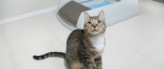
- Bring your cat for medical examination in a timely manner so as not to miss an incipient or progressing disease. Clinical examination usually includes standard blood and urine tests, ultrasound of the abdominal organs (blood tests and ultrasound are done on an empty stomach after 8-10 hours of fasting, but water should be left freely available), and if necessary, additional studies.
- If you notice any of the signs or a combination of them - frequent urge to urinate, blood in the urine, vocalization (the animal screams on the tray or simply calls you to pay close attention to it), urine in small quantities - you should contact a specialist as soon as possible.
- Prevention of stress - try to change the animal’s environment to a minimum (repairs, moving), give your cat as much of the required attention as possible, provide everything necessary for a comfortable environment (scratching posts, toys, beds), it is possible to use anti-stress medications (as prescribed by a doctor).
In our clinic you can receive the full range of services described. You can make an appointment through our call center 8(495)995-50-30, as well as through the feedback form on the website vetacademy.ru.
Take care of your four-legged friends!
Return to list
Treatment of urolithiasis in dogs
Therapeutic measures depend on the location of the urinary calculus, its chemical composition, as well as on the individual characteristics of the body. Most often it involves dissolving crystals or stones, and if there is no response to conservative treatment, surgical removal of large stones is used.
Before starting treatment, a comprehensive examination of the patient is carried out. It includes a clinical examination, clinical and biochemical blood tests, urine analysis, urine culture to determine the sensitivity of microflora to antibiotics, ultrasound, and x-ray to determine the size of large stones.