An X-ray of a cat or dog is performed in a veterinary clinic using special equipment. X-rays of the chest or other bones are used for suspected fractures, serious illnesses, and in other cases. The technique can be performed with contrast, with minimal impact on the cat’s body. The diagnostic procedure is carried out on kittens and adults only as prescribed by a veterinarian.
In what cases is it indicated?
A digital x-ray of the chest, posterior region or tail is performed in a veterinary clinic if indicated. Although the procedure is safe, it still has a slight effect on the condition of a healthy cat. It is worth examining the lungs, spine and other structures using x-rays in animals in the following cases:
- Mechanical damage. If a cat has a suspected bone or tail fracture, dislocation or joint injury, it is recommended to take an x-ray to confirm/refute the diagnosis.
- Respiratory diseases and inflammatory reactions in the trachea, bronchi or lungs.
- Falling from a window after a traffic accident.
- Malignant tumor in a cat of different localization.
- Impaired heart function.
- Suspicion of intestinal obstruction. The problem may be associated with a foreign object entering the gastrointestinal tract, as a result of which it is worth taking an x-ray of the stomach and other digestive organs.
- Enlargement or curvature of the pancreas.
- Formation of kidney stones.
- Disturbed structure of the urinary system organs.
- Suspicion of porto-systemic shunt. In a cat, the pathology is characterized by a congenital defect, as a result of which an extra vessel is present.
- During pregnancy in a cat. In a healthy individual, manipulation is performed to determine the number of kittens.
Veterinarians recommend X-raying a cat if spinal cord compression is suspected. In case of such a violation, such a diagnostic technique is the most informative.
How is the load reduced during x-rays?
All information about the radiation examinations performed, their number and radiation dose is entered into the medical record. If a critical dose accumulates over the course of a year, then prescribing another x-ray is highly undesirable.
To control the workload, the radiographer must have maximum information, so it is important to report all previous examinations and possible contraindications.
To protect the body, three main methods of protection are used:
- Protection by distance. The X-ray tube is placed in a special protective casing. It does not allow X-rays to pass through, which are directed at the patient through a special “window”. In addition, at the exit of the rays from the tube, an X-ray machine diaphragm is installed, with the help of which the irradiation field is increased or decreased.
- Time protection. The patient should be irradiated for as little time as possible (short shutter speeds when taking pictures), but not to the detriment of diagnostics. In this sense, images provide less radiation exposure than transillumination.
- Shielding protection. Parts of the body that are not to be photographed are covered with sheets and aprons-skirts made of leaded rubber. Particular attention is paid to the protection of the genital organs and thyroid gland, as they are the most sensitive to x-ray radiation.
Features of preparation
Before the barium test, the pet must first take Espumisan.
If an x-ray of a cat’s skull or other organs is done urgently, then no preparatory procedures are performed. If a diagnostic procedure with barium is planned in advance, then you need to know how to prepare your pet for it. The veterinarian can prescribe Espumisan to the cat, which is taken three times a day, 1-2 capsules. Such a preparatory measure before performing the manipulation is prescribed in the case of diagnosing the spinal column, bone structures of the pelvic floor, femur and humerus, as well as the abdominal cavity and lungs in cats. Preparation is required to reduce the accumulation of gases in different parts of the stomach and intestines. If there is excessive gas formation, the images may be inaccurate, causing the final results to be distorted. If an x-ray with a contrast agent is performed without anesthesia, it is recommended that the owners be present to fix the animal in the required position.
Preparing for a cat x-ray?
Filming is sometimes prescribed very urgently and urgently, if it is the consequences of a car accident, fall or other severe injury that occurred recently. In this case, of course, there can be no talk of any preparation; doctors work with what they have and try to draw a conclusion based on the results obtained.
How to prepare your pet for a cat x-ray procedure
If an x-ray is prescribed in advance, the doctor prescribes some medications
, which will help
make shooting
the sternum, abdomen, spine, hip, thigh and shoulder joints and parts
much cleaner
. The image will be clear and legible, which means the diagnosis becomes more correct. The course of treatment for your pet and its full recovery depend on this.
Next we will also mention how filming and x-rays actually happen. However, we must keep in mind and we will say more than once: the owners must be in the presence of the animal
. It is advisable that it is not one person, but at least two. They will fixate the pet while the photo is taken.
How do they do an X-ray of a cat?
Of course, X-raying cats is not so easy from the point of view that they are animals. Moreover, they are very capricious. So they strive to run away somewhere and interrupt the procedure. Moreover, if you simply try to force the animal to sit still, it takes a lot of time, plus you cannot take several x-rays in a row. Radiation does not cause harm several times a year, but if done more often, there will be some complications that can even develop quite serious.
It is for this reason that X-rays of cats are done using brute force, or rather with two or three people holding the animal in the position required for the photograph. It is advisable that these are the owners and owners of the cat. Or people whom the pet trusts and will not twist and turn in their hands. It is important to hold firmly enough
, because any movement can distort the results of the study.
If everything was done according to the rules, then the veterinarian, in the presence of the owner, within ten or fifteen minutes, will give a conclusion
and verdict on what was removed and received.
Our doctors are able to perform such a procedure at any time of the day, seven days a week
. If the animal is too restless, they often go so far as to use sedatives and drugs that help the cat relax. Such sacrifices have to be made for the sake of a good photo.
Sometimes the picture is taken only with the help of general anesthesia. Of course, anesthesia and anesthesia do not have a positive effect on any animals or even people. But recently, more and more often, drugs are appearing that are not completely harmless, but have fewer contraindications and consequences than their older counterparts. That is why you should not worry if the doctor prescribes this particular method of performing x-rays.
If such a method was prescribed, it was for one of the following reasons:
- X-ray for diagnosing pelvic or elbow joints.
- Images to determine osteochondritis of the shoulder.
- Knee joints, various injuries and ruptures associated with them.
- Various ugorgraphies, myelographies and portographies.
During the entire filming process, the animal is looked after by experienced veterinarians, anesthesiologists and part-time resuscitators. They also observe the animal after the shooting, because animals experience recovery from anesthesia quite hard. Just like people, actually. To carry out the shooting and the whole process in general, the conclusions and consultations of a therapist , orthopedist-tramatologist and neurologist . The combination of these three veterinary areas will provide a complete guarantee that the procedure is necessary and the cat is able to tolerate anesthesia and filming even with its individual characteristics of the body.
How do they do it?
Since taking x-rays requires the use of special equipment, it cannot be performed at home; the procedure is performed exclusively in a veterinary clinic. In rare cases, x-rays of a cat's paw or other areas of the body are performed under local anesthesia. Most often, 2-3 people are involved in performing the manipulation, who could provide the pet with a motionless body position so that the specialist can take high-quality photographs. The duration of the diagnostic procedure is no more than a quarter of an hour, and the cat does not experience pain or other unpleasant sensations when performing an x-ray. The advantage of this diagnostic technique is the ability to carry it out at any time without prior preparation. For a more accurate clinical picture of the pathology, the pet is often given barium sulfate to drink. This procedure is often performed when examining the stomach for the presence of a foreign body. X-ray is performed as follows:
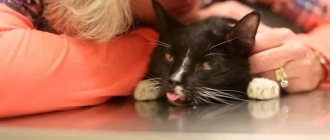
First, the animal must be laid down and secured on the table.
- The owners fix the cat on a special table. Some veterinary clinics provide special protective equipment (aprons or collars) so that owners remain clean during the procedure. If the doctor deems it necessary, the x-ray is performed under local anesthesia.
- The specialist sets up the x-ray and takes several pictures. For injuries, a direct, posterior or lateral projection is performed.
- The cat is released and the veterinarian deciphers the results of the diagnostic examination to the owners. The specialist determines how far the indicators are from the norm and, if necessary, prescribes treatment.
X-ray of a cat with a foreign body in the stomach or intestines
Intestinal obstruction is very dangerous for the body, so if you discover that the cat has swallowed any object, you should immediately contact a veterinary clinic.
Reasons for ingesting various substances:
- Games during which the cat accidentally swallows a toy;
- Accumulations of hair in the stomach;
- Chewing rustling things (candy wrappers, bags, tinsel, etc.);
- Feeding bones, including fish bones.
extracted propylene thread
Swallowed objects remain in the stomach, blocking the exit to the intestines, or completely stop the stomach from working. If a cat swallows a fish bone, it can injure the stomach or intestinal walls. When a cat swallows threads or tinsel, they are placed along a significant gap, thereby collecting intestinal loops, resulting in injury to its walls. If there is thread or tinsel sticking out of your cat's anus, you should never pull it out yourself. This can lead to serious consequences.
Symptoms of a foreign body in a cat’s body:
- Refusal to eat;
- Lack of stool;
- Lethargy;
- In some cases, you can feel a hard object in the abdominal cavity.
To determine the exact position of a foreign body in a cat's body, radiography is used. The photographs show the location and size of the object. X-ray - the picture is taken in lateral and frontal projections, sometimes a barium solution is used for a more accurate image. For a more detailed study, the picture is taken in horizontal and vertical positions of the animal.
Is the procedure harmful?
Some owners believe that X-raying the head of a cat or dog is not safe and has a negative impact on the health of the pet. This information is untrue, since during manipulation the radiation occurs in a minimal amount, which does not in any way affect the condition of the pet. In addition, a disease that was not diagnosed in time using x-rays or other diagnostic methods can pose a greater danger than the influence of x-rays.
Modern veterinary clinics use special X-ray equipment to protect the cat from scattered radiation.
How many times can an x-ray be taken?
If we are talking about analog devices, then experts recommend a break between irradiations of 3 weeks and take one photo per visit.
. However, it happens that it is necessary to increase the number of studies, then they are carried out every couple of days, reducing the negative impact as much as possible. Several x-rays on an analog device in one day can have a bad effect on your health.
The invention of digital equipment has made it possible to greatly reduce risks and allow for more frequent x-ray examinations. There is no longer any need to make compromises between harm and health benefits; doctors prescribe as many procedures as necessary to effectively monitor the progress of treatment.
Benefits of manipulation
It is not for nothing that such a diagnostic technique for any cat pathology is one of the most popular, since it has a lot of positive aspects. With an x-ray, it is possible to separate the image and identify pathology in the lower and upper areas, while it is possible to zoom in on the image in order to more accurately examine the pathological areas. It is also possible to make a recording in real time, which will display the work of internal organs. Based on the X-ray results, the specialist assesses the clinical picture as accurately as possible and selects individual therapy for the cat.
Why choose fluoroscopy at the RosVet VC?
In an emergency situation, specialists of the RosVet VC advise calling +7. The clinic receives visitors 24 hours a day, so urgent fluoroscopy can be done around the clock, as well as submitting a request for a routine examination, or as prescribed by a veterinarian during an examination.
Our veterinary center employs experienced radiologists who are able to interpret images with accuracy that does not cast doubt on the final diagnosis. The procedure itself is carried out with minimal stress for the pet, regardless of the required projection. If necessary, the animal is given sedation (anesthesia).
If you need to have your pet x-rayed, please contact the RosVet VC by phone: +7(495) 256-11-11. Applications are accepted around the clock. In case of urgent need, the clinic is available 24 hours a day.
Can X-ray examinations be performed on pregnant women?
In the first half of the term, the study is done only according to strict indications. In the second half of the term, you can do as much research as you like.
Another interesting fact. People often ask X-ray technicians to wear more protective lead aprons. Let's say right away that this is useless. You receive the same dose of radiation from covered and exposed parts of your body.
And remember that a doctor will never order an x-ray just out of curiosity. X-ray is one of the types of diagnostics of the area of proposed treatment and the anatomical features of the oral cavity.
Come to our clinic and we will make every effort to make your stay comfortable and enjoyable!
Good to know
- Thoracic radiography
- The role of the Buchanan coefficient in assessing heart size in dogs and cats
- The role of the cardiovertebral index in radiographic assessment of heart size in dogs and cats
- The role of the cardiothoracic (cardiothoracic) index in the diagnosis of cardiomegaly using a thoracic radiograph performed in a direct ventrodorsal projection
- Methods for visual assessment of the shape and size of the heart
- How long can a dog with heart failure live?
- Basioapicosternal angle in dogs and cats
- Vertebrotracheal angle (tracheovertebral angle) in dogs and cats
- Peculiarities of interpretation of chest radiography indicators in obesity
- Protocol for radiographic examination of the heart of dogs and cats
What equipment is used
To carry out 3D diagnostics, a three-dimensional computed tomograph SOREDEX Scanora 3D with advanced functionality is used. This is the latest generation equipment, which allows you to obtain three-dimensional images of the anatomical structures of the maxillofacial region in a few seconds, with the least radiation exposure for the patient.
The program analyzes the obtained multiplanar sections and builds them into a 3D model, thanks to which the specialist is able to accurately assess the condition of the dental system, detect all pathological processes occurring in this area and competently plan a treatment regimen.
A virtual 3-dimensional model of the scanned area can be recorded on any digital media (CD, flash drive), which allows the attending physician, if necessary, to view diagnostic data or involve related specialists in the analysis of the received information.