Features of changing baby teeth in kittens
Thanks to its teeth, a cat holds prey in its mouth, carries kittens, and defends itself when danger arises. The front teeth help cats wash themselves and chew out tangles and stuck debris. Since the change of teeth is an important stage in the growing up of an animal, you need to be aware of how the change of baby teeth and the eruption of permanent molars occur: by what symptoms can you understand that your pet is going through this stage and how to make it comfortable for your pet.
Milk teeth in kittens
Kittens are born completely toothless. The first milk teeth erupt at the age of two weeks and differ from permanent teeth in their specific structure.
Structural features and differences from permanent teeth
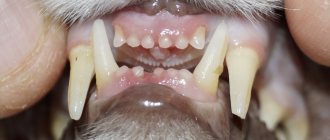
Milk teeth in kittens are straight (in adult cats they are powerful and slightly curved) and have a milky white color (hence the name - milk teeth). In addition, they are sharp, like small needles.
Characteristic features are the primary canines, which erupt by the first month and change closer to five months. They are very thin and curved. In addition, on the inside of each fang there is a clearly visible second tooth, which falls out after the change.
In total, the kitten has twenty-six milk teeth. All of them should be fully formed by the age of two months: twelve incisors, four canines and ten small molars. Below is the dental formula of baby teeth.
For simplicity, veterinarians use dental formulas: incisors are designated by the letter I, canines - C, premolars - P, molars - M
If several or even one tooth is missing by the specified age, you should seek help from a veterinarian, since this problem may become a symptom of a more serious disease.
Table: timing of baby teeth eruption in kittens
Formation of teeth in a kitten
The structure of the teeth of cats is typical for their species, and zoologists consider the structure of the dental system as its morphological feature. The structure of the dental system reflects the nature of the diet, and the cat is an undisputed predator - all its dental crowns are conical in shape and are designed to tear hunted game into pieces. The teeth located on the jaw form the upper and lower arcades, respectively. To form a breed characteristic—the appearance of a cat’s face—the correct formation of dental arcades is necessary. Teeth are necessary for capturing, holding and tearing prey; the cat uses them to intimidate the enemy, in fights, for grooming, and for carrying small objects, including kittens, in its mouth.
The formation of tooth buds in cats occurs during embryonic development in the form of a dental plate - this is a fold of epithelium containing tooth buds. The inner layer of cells of the dental plate - anameloblasts, form tooth enamel; The cells of the outer layer are odontoblasts, they form dentin. The connective tissue surrounding the tooth germ forms cement.
The development of teeth is associated with their eruption, when the teeth come out through the mucous membrane of the gums.
Teeth are made up of tissues:
- dentin is the modified bone tissue that makes up most of the tooth. Dentin forms the tooth cavity, where the pulp containing blood vessels and nerve fibers is located;
- enamel is a very durable white tissue that covers the surface of the tooth from the outside, where it comes into contact with the external environment;
- cement - covers the parts of the tooth located in its alveolus - the cell of the jaw where the tooth is located. Cementum is represented by bone tissue of a layered structure.
The following parts can be distinguished in the appearance of a tooth:
- tooth crown - part of the tooth that protrudes freely into the oral cavity and has a chewing surface;
- tooth root - located in the alveolus; the cementum of the tooth root and the periosteum of the alveoli are connected by the periodontium;
- neck of the tooth - connects the crown of the tooth with its root, here the coating of the tooth changes, the enamel of the crown passes into the cement of the tooth root. The neck of the tooth is normally located in the gum.
Based on function and form, they are distinguished:
- incisors - small, flat-shaped teeth located on the jaws in front and having one root. The incisor crown has three projections that wear away over time; The cat uses the incisors for grasping and holding, as well as grooming; the roots of the incisors are shallowly embedded in the jaw, so these teeth are often lost with age;
- fangs - the cat’s fangs are dagger-shaped, they are sharp and long, their roots are immersed very deeply in the bone tissue of the jaws; used to kill prey; the cat uses its fangs to separate the cervical vertebrae of the victim - this is a characteristic hunting feature of small cats; fangs are used for tearing food; each fang has only one root, the fangs are located next to the incisors;
- premolars - small molars; Kittens also have them; premolars are needed for grinding food and have a chopping effect; the number of premolars is different on the upper and lower jaws: there are 6 on top, 4 on the bottom. They have 2 or 3 roots, so before removing a premolar, the veterinarian prescribes an x-ray to clarify the number of roots and not leave one of them in the jaw; located next to the fangs;
- molars - large molars - do not have their milk equivalent, they grow immediately in the form of permanent teeth; molars are also designed for cutting and crushing food; located at the edges of the dental arcades; The upper molars have one root each, the lower molars have 2.
During the teething period, cats try everything, it is especially dangerous when they chew wires
When and in what quantity do baby teeth appear?
At birth, a kitten has no teeth at all, but starting from the 2nd week of life, milk teeth begin to appear. A cat's primary set of teeth is incomplete because molars, the large molars, are missing. They are called milk teeth because they appear when the kitten feeds on milk, and also because of the specific color of the tooth enamel - milky white, translucent. Baby teeth are smaller, sharper and more fragile compared to permanent teeth. In total, the milk set contains 26 teeth: 12 incisors, 4 canines, 10 premolars.
Timing for the eruption of baby teeth:
- incisors: the first primary incisor erupts at 2–3 weeks; the second - at 2.5–4 weeks; third - at 3–4 weeks;
- fangs: milk fangs erupt at 3–4 weeks of a kitten’s life;
- premolars: eruption in 4 to 8 weeks.
It is generally accepted that the age of a kitten can be accurately determined by its teeth, but this is not entirely true, since individual and breed characteristics of the timing of teething can create a large error in the accuracy of this method.
The practical benefit of knowing the average timing of the eruption of baby teeth is expressed in the ability to control their development, since the development of teeth is also an indicator of the development of the kitten.
The eruption of baby teeth usually goes unnoticed by the kitten’s owner, because the cat is still too small.
By 2 months the kitten has a full set of baby teeth.
At what age does teeth change?
The replacement of baby teeth with permanent ones occurs when the kitten is 3–6 months old. The permanent set includes 30 teeth: 12 incisors, 4 canines, 10 premolars, 4 molars.
Changing baby teeth to permanent ones
At the age of three months, kittens begin to lose their milk teeth, gradually being replaced by molars . This process is usually complete by six or seven months, but don't worry if it takes longer for your kitten. Such deviations are usually associated with the breed of cat or characteristics of individual development.
The order of changing teeth should not be disturbed. The first to be replaced are the incisors, then the canines, and lastly the molars and premolars are replaced.
However, if a new tooth grows on a baby tooth and this causes discomfort to the pet, then you should definitely consult a specialist. If such growth does not bother the animal, then a visit to the veterinarian can be postponed, since baby teeth can calmly and painlessly fall out even after the main shift.
Teeth change schedule
A healthy adult cat should have thirty permanent teeth: twelve incisors, four canines, ten premolars and four molars. Each of them serves her for a specific purpose, for example, incisors are needed to tear food, fangs help to capture prey, and so on.
A cat's permanent dental formula is formed after six months. It includes: on top - three incisors, one canine, three premolars, one molar; below - three incisors, one canine, two premolars, one molar. When calculating, all coefficients are doubled, so the total is thirty permanent teeth.
This is what the jaw of an adult healthy cat should look like with timely and correct teeth replacement.
Table: schedule for the eruption of permanent teeth and their functions
Symptoms of tooth change
The process of changing teeth can begin and even end unnoticed by you, since the kitten usually does not experience pain. Most often, the change of teeth becomes obvious when a lost baby tooth is found.
Nevertheless, there are a number of symptoms that will help you navigate and notice in time the process of changing teeth:
- When a kitten's teeth change, an unpleasant odor may appear from the mouth, which is often associated with poor nutrition. There is no need to do anything about this, it will quickly disappear once the teeth renewal process is completed.
- Kittens may experience discomfort during the actual cutting of new teeth, so the animal's behavior will change slightly. For example, a slight increase in temperature is likely (the norm is a temperature of 38 ° C to 39 ° C, for small kittens a higher temperature of up to 39.5 ° C is typical). As a result, the kitten feels the cold more strongly and tries to spend more time next to the warm body of the owner: on the lap or in the arms. At night, pets can crawl under the covers, even if this was previously uncharacteristic for them.
If your pet suddenly loves lying under a blanket, this may be a sign of discomfort when changing teeth.
Be very careful that your kitten does not start chewing wires or other objects that could harm him.
The author of these lines recently encountered the process of changing teeth in his kitten. Each animal exhibits specific symptoms, and when problems arise, it is easy for owners who know their animals to notice changes in behavior. So, my kitten usually did not hide under the blanket and did not play with some toys. When his teeth changed, he constantly chewed rubber sticks and tried to bite the scratching post. In this case, the baby teeth did not fall out immediately, but after the fangs were replaced.
Video: kitten teeth falling out
Caring for a kitten during the period of teeth change
The period of changing teeth can really become a test for an inexperienced owner. However, it is necessary to understand that the animal is not sick, does not need to take medications and constant visits from veterinarians. Most often, for a cat, everything happens, if not unnoticed, then at least painlessly.
There is no need to allow the kitten to chew furniture, spoil things, and especially not to bite and scratch you. In the future, this habit could become a really serious problem. Changing teeth is a natural process for an animal; therefore, the initially established rules of behavior must be preserved.
However, the task of every responsible owner is to make this period easier for the animal, take care of proper nutrition, brushing teeth and the availability of special toys.
Special food
A kitten needs proper and complete nutrition . However, despite the abundance of food created specifically for the period of teeth change, the animal may react negatively to a sudden change in food. Therefore, you need to ensure that your pet has the necessary vitamins and supplements in its diet, thanks to which permanent teeth will grow strong.
There are nutritional principles that must be followed when a kitten’s teeth change:
- First of all, you need to avoid overly soft food: kittens can swallow lost teeth along with soft food, which can lead to damage to the esophagus. The food should be large and slightly rough. Regular dry food for kittens is ideal.
Dry food is the best option during teeth change
Bones are an important part of the diet and serve to prevent the kitten from chewing your things.
Veterinarians advise not to mix natural food (meat, fish, vegetables, etc.) and dry food. If you switched your kitten to dry food at an early age, then you should stick to it. The fact is that these types of food are digested differently, and when mixed, the kitten may experience bloating or even colic.
When choosing a feeding method, you should consult a veterinarian: each pet is individual, many may not be suitable for dry or natural food
Vaccination during the period of teeth change
Very often, owners are faced with an important question: is it possible to get vaccinations or other vaccinations while changing teeth? Veterinarians speak out unequivocally - changing teeth is already a serious burden for a kitten. Against the backdrop of fever and pain, getting vaccinated can be dangerous. This not only adversely affects the kitten’s immune system, but also leads to stunted growth of the animal.
Vaccination during the period of teeth change is not recommended.
It is important to follow the vaccination schedule for your pets as prescribed by your doctor. The vaccination schedule is set individually, taking into account the age characteristics of the body and physiological state.
My kitten was scheduled for the first vaccination very late. This was due to the fact that he was found on the street and many tests had to be done to assess his health. The time for his vaccination was due to change his teeth, so it had to be postponed for a while (until the temperature passed and slight inflammation of the gums subsided).
Teeth cleaning
Many owners ignore brushing their teeth. However, constant oral care will help keep your animal's teeth strong. It is necessary to accustom your cat to brushing its teeth from the early months of life so that in the future it will adequately perceive this procedure. If the cat refuses to accept traditional brushing with a toothbrush or powder, pet stores sell special gels for disinfecting the oral cavity. You can combine them with special foods and vitamins that include coarse fibers. It is recommended to brush your teeth once every three to four weeks.
Brushing your teeth is an important procedure that your kitten needs to be accustomed to.
During the change of teeth, kittens' gums can become inflamed, so you need to purchase a gel that also contains an anesthetic and anti-inflammatory, which can greatly alleviate your pet's condition.
Possible complications during the period of teeth change
Changing teeth is a natural process that can occur without problems for the animal. However, during the period of active growth of molars, you should inspect the animal’s mouth at least once every few days. It is important to consider that reddish and slightly inflamed gums are normal. However, in some cases complications arise. A caring owner should notice the problem in time and be sure to contact a veterinarian.
The most common complications that are visible to the naked eye:
- Suppuration of the wound at the site of the lost tooth.
- The animal shows anxiety, meows pitifully, and may experience severe lethargy.
- The kitten refuses to eat for two days.
- Very severe inflammation of the gums.
- Wounds appear from a missing milk tooth, in the place of which a permanent tooth has already grown.
- Some of the baby teeth have not fallen out, although the permanent ones have already grown in and the time for changing teeth has already passed.
In these cases, you should take the kitten to the clinic or call a doctor at home.
Don't hesitate to contact your veterinarian, even if your pet doesn't show signs of restlessness, lethargy, etc. A healthy animal also needs control, so veterinarians are very loyal to owners who bring healthy animals for examination and ask to monitor the process of changing teeth.
Gum inflammation
Inflammation of the gums is one of the most common complications during the period of teeth change. A little inflammation is normal, but in some cases it lingers and the gums become red.
Inflamed gums become swollen and red
Signs of gum inflammation:
- the animal is restless;
- refuses to eat due to painful sensations;
- tries to chew more;
- rubs its muzzle against everything in an attempt to relieve pain;
- profuse salivation occurs;
- swelling and intense redness are noticeable.
Of course, it is better to have a veterinarian do the diagnosis and treatment, but gum inflammation may go away when the kitten is switched to softer food.
“Stuck” baby teeth: signs and treatment
A more serious problem is the residual baby teeth, which do not fall out until the permanent molar emerges from the gum. Due to improper growth of molars, the bite may be disrupted, which will lead to high trauma to the cat’s gums, cheeks and lips.
If a baby tooth has not fallen out, but a molar has already grown in its place, you should consult a doctor, as this can lead to malocclusion
Baby teeth become a problem if:
- the kitten has not lost some of its milk teeth after six months;
- there are loose baby teeth with active growth of the molars underneath them.
Unlike sore gums, stuck baby teeth should only be treated by a veterinarian. Unfortunately, this problem is often solved only by surgery under anesthesia, since it is impossible for baby teeth to fall out on their own.
The kitten constantly demands attention. From the very first days until old age, your animal will need constant care. However, in the life of every pet there are certain periods that will require even more care from you. One of these periods is the process of changing teeth, which marks the beginning of adulthood in a cat. Therefore, it is important to know how it happens, how to care for a kitten and what to do if health problems arise.
Causes of loss of fangs
Loss of teeth, including fangs, can have several causes:
- physiological – natural change of baby teeth to permanent ones;
- age-related – tooth loss caused by degenerative processes in the aging body;
- pathological – tooth loss occurs as a result of pathology of the tooth crown, gums or trauma.
Natural change of teeth is a temporary process. In the wild, the female helps feed during this period.
Physiological change of teeth
Kittens are born toothless and feed on their mother's milk until their first teeth appear. First, the incisors erupt, and this happens at the age of 1-2 weeks. Then after 3-4 weeks the canines appear and after 6-8 weeks the premolar teeth finish erupting. Babies have no molars. By 2 months, the kitten should have 26 baby teeth. They are whiter and thinner than permanent ones.
The replacement of baby teeth with permanent ones begins at the age of 3.5-5.5 months. The procedure is the same as for teething:
- 3.5-5.5 months. – incisors;
- 5.5-6.5 months. – fangs;
- 4.0-5.0 months. – pre-radical.
Molars appear in babies at the age of 5-6 months. By 7 months, the physiological change of teeth should be completely completed. But in some cases it drags on for up to 9 months.
In females, the replacement of milk teeth begins later than in males.
An adult cat's complete dental formula is:
- 12 incisors - 6 pieces each on the lower and upper jaws;
- 4 fangs;
- 10 premolars (premolars);
- 4 molars.
There are a total of 30 permanent teeth in a cat's mouth. Signs of tooth change are:
- hypersalivation - excessive salivation;
- the desire to chew and gnaw inedible objects;
- decreased appetite;
- mood changes – irritability, aggressiveness, increased excitability;
- decreased physical activity due to malnutrition.
You need to help your baby get rid of a loose baby tooth only if it interferes with the eruption of a permanent one. Sometimes instead of 4 fangs there are 8 – the milk ones have not yet fallen out, but the permanent ones have already appeared. This happens due to the fact that fangs are formed in separate follicles. Normally, the kitten will get rid of the tooth that is causing discomfort on its own, but the help of a veterinarian may be required.
Age-related canine loss
With age, degenerative processes occur in the animal’s body:
- mineral metabolism is disrupted - teeth become less durable, they wear out and even break. The longest ones in the jaw, the canines, are especially affected;
- Collagen synthesis decreases - teeth begin to loosen due to weakening of retaining fibers and loosening of gum tissue;
- Immunity decreases, which increases the risk of infections and the development of dental pathologies.
Signs of wear on dental crowns (the part of the tooth that protrudes above the gum) can be seen in cats aged 3-5 years. The animal's teeth become dull and darker due to deposits of tartar and soft plaque.
Cats older than 5-6 years may already be missing some teeth. To extend the life of the dental system of a domestic predator, you need to monitor oral hygiene from the first months of life.
Even with the complete loss of fangs and significant thinning of the dentition, a domestic cat will not suffer from hunger. Companies involved in the production of pet food produce a number of special lines of food for cats older than 7-12 years.
Pathological tooth loss
Dental problems can lead to the loss of canines and other teeth, even in a young animal. Caries is a fairly rare pathology in cats, which is due to the structure of the predator’s dental crown. Fangs are least often affected by caries, since they do not have grooves and depressions on their surface in which food can accumulate. But other pathologies can lead to caries:
- traumatic chipping of fangs as a result of gnawing hard objects;
- trauma to the gums by sharp fish and poultry bones with subsequent penetration of infection into the wounds;
- metabolic disorders leading to thinning of the enamel;
- lack of minerals and vitamins in the diet necessary for the mineralization of teeth;
- deposits of tartar, which is a nutrient substrate for pathogens.
In addition to caries, a cat can lose its fangs as a result of:
- periodontitis;
- gingivitis;
- odontogenic osteomyelitis;
- tartar;
- pulpitis.
Pets suffer from the same dental problems as their owners. A cat can lose its fangs from mechanical damage or injury - breaking them in a fight, falling from a height, or a direct blow to the face.
Today there are not many veterinary clinics that offer a full range of services for treating dental diseases of pets. But in large cities, a furry pet can have an implant installed in place of a lost canine or premolar. The question is the cost of this service. The high cost significantly reduces the demand for dental prosthetics in cats.
Changing teeth in cats
Kittens, like other pets, are born without teeth. As kittens grow older, they develop baby teeth one after another, which are eventually replaced by permanent teeth. The process of the appearance and change of teeth usually goes unnoticed for owners, as it usually takes place without any complications.
Knowledge of the process of changing baby teeth to permanent teeth will allow kitten owners to promptly notice and eliminate possible problems in the kittens’ oral cavity.
Formation of a cat's dental bite.
A kitten's milk teeth begin to erupt from the 2nd week of life and finish erupting by 6-8 weeks of age. In total, by the age of 2 months, a kitten has 26 milk teeth, which form the cat’s bite.
The order of eruption of baby teeth in a cat.
The kitten's incisors are the first to erupt at 2-4 weeks of age. At 3-4 weeks of age, fangs appear. Premolars appear in a kitten at the age of 6-8 weeks.
A kitten's emerging baby teeth are thinner than their permanent teeth. The baby teeth will serve the kitten for several months. If the cat has enough milk, and the kittens are given balanced supplementary feeding in a timely manner, there is no lag in weight gain and development, the replacement of milk teeth with permanent teeth will begin at the age of 3–4 months.
How do baby teeth change to permanent teeth?.
The replacement of baby teeth with permanent teeth in a kitten begins at 3-5 months of age and ends at 7-8 months, when 30 permanent teeth form a permanent molar bite. Constant contact and mechanical irritation causes destruction of the roots of baby teeth, which stimulates loosening and their loss.
The process of replacing baby teeth with permanent teeth is usually painless and goes unnoticed for cat owners.
The resulting permanent dentition in a cat is represented by:
- 12 incisors (6 incisors each on the lower and upper jaws).
- 4 fangs (two on each jaw, running along the edges of the incisors).
- 10 premolars (start behind the canines, with 6 premolars in the upper jaw and 4 premolars in the lower jaw).
- 4 molars (2 on each jaw). These 4 molars in the cat are missing in the primary dentition.
The order of replacement of baby teeth with permanent teeth.
The replacement of milk teeth with permanent ones in a cat occurs in the same order as the appearance of milk teeth.
- First, at 4-5 months, the incisors are replaced.
- Then, at 4-6 months, the fangs are replaced.
- At 5-6 months, the premolars are replaced.
- At the end of the 6th month, molars grow.
Correction of canine position disorders in dogs and cats
F. Genne
Violation of the position of the canines is a change in the bite, which affects their functional state.
Incorrect canine alignment can be caused by a misalignment of the primary teeth or an abnormality in the size of the mandible.
The categories of these deviations from the norm are determined by the orientation of the canines in space.
The first part of the article presents a classification of violations of the position of the canines, while the second discusses the issue of ways to eliminate defects.
Classification
Abnormalities in the position of the teeth In the transverse projection An anomaly in the position of the fangs is much more often noted in the transverse projection. It is mainly found at the level of the lower canines, the position of which has a pronounced direction towards the tongue.
This anomaly, called canine convergence, refers to linguoversion (severe inclination of the canines towards the tongue) or linguoposition (severe displacement of dental implants towards the tongue). The etiology of these dental disorders is not well understood. If in small breeds of animals the persistence of primary canines is the main factor of these disorders, then in others this cause of defect is encountered much less frequently. Also, the lower canines may have a pronounced inclination in the lateral direction (vestibular version), which is also called divergence.
As for the upper canines, violation of their position is much less common. We can observe their deviation towards the palate (due to the persistence of baby teeth).
In lateral projection In dogs and cats, rostral deviation (rostroversion) of the upper canines can also be observed. This is most often observed in small breeds of dogs (Yorkshire terrier, poodle, dwarf dachshund, etc.) due to the persistence of primary canines. In addition, depending on the breed, the persistence of primary canines causes a more or less pronounced disturbance in the position of the permanent canines. This feature is typical for Italian and Scottish Italian greyhounds (photo 1). In any case, particularly in these breeds, it is very difficult to establish whether the persistence of the primary canine is the cause of this type of disorder or whether it provokes rostroversion of the primary canine.
| Photo 1. Rostroversion of the upper canine in an Italian greyhound. A persistent milk tooth was recently removed (the wound caudal to the tooth), but a fragment remained. |
In cats of brachycephalic breeds (Persians, exotics), these disorders are of a specific nature. If there is rostrodeviation of the upper canines in association with the persistence of primary teeth, then it should be borne in mind that one of the reasons for the intensity of this pathology is the maxillary-facial modification that we observe in brachycephalic breeds. As for the lower canines, the violation of their position is caused by the rostral deviation of the upper canine or a skeletal anomaly, characteristic of brachycephals.
Vertical projection A fairly common anomaly in the position of the canines in the vertical projection is egression (progression of one or more teeth that do not have antagonists), and it is observed mainly in small breeds of dogs. Egression is also noted with malocclusion.
Violation of the position of the teeth associated with changes in the structure of the skeleton In the transverse projection As for the violation of the position of the main teeth at the level of the lower canines (“convergent canines”), it is recommended to introduce the term mandibular endognathium (narrowing of the lower jaw). This skeletal anomaly in transverse view corresponds to a pronounced "internal" displacement of the position of the canines in relation to the maxilla.
In the lateral projection In the case of upper or lower prognathism (or brachygnathism), a violation of the position of the upper canines in relation to the lower canines is noted, due to the shortening of one of the jaws.
These two anomalies are regrouped because they are predominantly volumetric anomalies (transverse and sagittal projection), in which the mandible is narrower or shorter (micromandibularia). In this case, the lower canines have a more internal and caudal position.
Guide to action
Reason for consultation and medical history There are two reasons for consulting a doctor:
- mainly rostral deviation or divergence of the lower canines (the aesthetic factor plays a role here);
- indications for treatment in case of anomaly of the lower canines in the transverse projection (convergent canines) or in the case of rostroversion of the upper canines or micromandibulia.
The main causative factors that must be taken into account are: breed, how long ago the anomaly occurred, the state of occlusion due to the replacement or spontaneous loss of primary teeth, the presence of primary teeth, as well as the presence of anomalies during the growth period.
Determining the severity of the violation The main significance is the violation of the position of the lower canines. Due to their size, changes in the position of these teeth, mainly in the transverse projection, negatively affect the condition of the gums or palate. Sometimes the disease develops before the change of baby teeth, but becomes more pronounced from the moment the permanent teeth appear (from the age of five months). Treatment should be carried out before the age of one year (photo 5, 6).
| Photo 5. Damage to the palate due to linguoversion of the lower canine in a puppy aged six months. It should be noted that the canines have not yet fully erupted. | Photo 6. Occlusion of the maxillary teeth in this dog indicates damage to the palate, which is localized in the space between the teeth. Effective orthodontic treatment was carried out (Fig. 1). The lips fit tightly to the upper canine, which ensures fixation of the removable appliance. |
| Figure 1. Treatment options for palatal injuries caused by mandibular canine malalignment. 1) – damage to the palate is noted in the sagittal plane in relation to the space between the angle of the canine and the first third of its width. Orthodontic treatment is required. 2) – damage to the palate, located in the sagittal projection at the level of the second third of the width of the canine. It is necessary to amputate the crown. 3) – damage to the palate, localized in the sagittal projection at the level of the third third of the width of the canine. It is necessary to carry out amputation of the tooth crown or orthodontic treatment aimed at repositioning the tooth caudal to the upper canine, the tooth should be located between this canine and the first premolar |
Rostral deviation of the upper canines provokes a more or less complete closure of the space between the teeth (forming the shape of an angle) into which the lower canines lie. Consequently, the lower canine, subject as a result of the above changes to a certain degree of lateral (vestibuloversion) and/or rostral (rostroversion) deviation, provokes injury to the upper lip or covers it.
Making a diagnosis At the beginning of the consultation, it is necessary to carry out a differential diagnosis, if possible, between skeletal anomalies and a violation of the position of the tooth.
An external clinical examination of the patient allows one to assess the degree of symmetry of the skull and facial surface of the head, as well as the position and shape of the main elements of the skeleton.
An oral cavity examination allows you to evaluate:
• occlusion of incisors, canines and premolars; • position of premolars (overfilling, rotation or displacement); • the shape of the mandible (straightness and parallelism in terms of occlusion or curvature with an open bite or gaping at the premolar level).
Before starting treatment, it is necessary to very carefully and completely examine the oral cavity under general anesthesia, which also makes it possible to conduct radiographic examinations inside the oral cavity. Radiography makes it possible to identify remaining fragments of the roots of primary teeth (photo 2), as well as to assess the stage of development of the roots of the corresponding teeth.
| Photo 2. Rostroversion of the upper canine in an Italian greyhound at the age of six months. The tooth is immature: the pulp canal is very wide, the root wall is thin, and the apex is unformed. It should be noted the persistence of a fragment of the root of a primary tooth (arrow). This persistence distorts the position of the main tooth. |
Treatment
Lower canines Lingual deviation of the lower canines Lingual deviation of the lower canines is most often accompanied by disturbances in the position of the teeth (linguoversion or linguoposition), the orthodontic treatment of which is quite well developed.
If baby teeth are still present, it is recommended to remove them immediately with care so as not to damage the root. In case of doubt, X-rays are used.
The decision for orthodontic treatment can be made when the lower canines have grown to half their height, which corresponds to the animal's age of approximately six months. The therapeutic option (choice), depending on the situation, is to install an active appliance between the lower canines or a passive one, which is also called functional, used on the upper jaw (at an angle).
If treatment is carried out before the full growth of the canines (at the age of 7 months), it is recommended to use:
• active flexible, usually hinged apparatus, so as not to change the direction of divergent growth of the lower canines; • functional appliance (angled), removable, designed for the upper jaw, which does not put pressure on the lower canines.
In addition, the transverse growth of the maxilla should not be inhibited during this period, which requires the use of an inclined hinged or “telescopic” appliance that meets these requirements (photo 7, 8). In adults it can be used with a fixed inclination.
| Photo 7. Installation of a functional removable appliance (inclined). The device is held passively using the lips. | Photo 8. The result of orthodontic correction after removal of the apparatus. |
Lingual and caudal deviation of the lower canines • Orthodontic treatment If the anomaly seen in the sagittal projection (caudal deviation) is not so pronounced, then a functional appliance (at an angle) can be used to correct the tooth(s) while simultaneously performing rostroversion (moving forward) or vestibuloversion (lateral displacement). During the examination, we can determine the boundary of the first third of the length of the upper canine (photo 6). The lower canines, which extend beyond its limits when the jaws are clenched (Figure 1, Photo 8), should be treated primarily in a different way: with the help of an active, more complex orthodontic apparatus (Photos 9, 10) or by amputation of the crown.
| Photo 9. Damage to the palate in the sagittal plane, at the level of the second third of the width of the canine. The most adapted treatment is crown amputation | Photo 10. Linguoversion and distoposition (more caudal position) of the lower canine. |
• Crown amputation Endodontic therapy is a priority when orthodontic treatment is contraindicated or undesirable. It allows you to avoid injury to the palate caused by the fangs if they are incorrectly positioned (photo 4, 8). This method of treatment is resorted to when the animals are over seven months old, because this corresponds to the period of formation of secondary, that is, more durable dentin at the crown level. This treatment is based on amputation of the upper part of the crown (approximately half). The tooth is then subjected to partial devitalization by eliminating the pulp (pulpotomy or partial pulpectomy). As a result of the manipulation, the formation of secondary dentin in the tooth crown is suspended. And vice versa, in its radicular (root) part the pulp is preserved: for this purpose we place a biological coating (“pulp combing”), which allows us to maintain its vital activity (normal root maturation) and in particular the closure of the apex (apexogenesis) and thickening of the root part of the wall tooth (secondary dentinogenesis). X-ray control for an objective assessment of its physiological formation is carried out after four or six months (photo 11, 12).
| Photo 11. Correction carried out by installing the maxillary and mandibular active apparatus. | Photo 12. Amputation of the crown with pulpotomy and “combing” of the pulp in a puppy at the age of seven months. The root is not yet ripe. Pulp brushing material and obturation are recognized by detecting a radiopaque mass. |
Rostral and vestibular deviation of the lower canines In case of deviation of one or two lower canines outward (lateral) and forward (rostral), the upper lip is captured, which is the reason for consultation. If the deviation is mild, then the canine(s) may rest against the upper lip.
This positional imbalance, either unilateral or bilateral, is common in brachycephalic cat breeds in association with skeletal disharmony. In this case, orthodontic treatment is not recommended, since the disorder occurs not at the level of the teeth, but at the skeleton. In cases where a violation of the position of the teeth is not associated with a skeletal abnormality, it is often unilateral (one-sided) in nature. The lower canine may be in a state of rostroversion due to that of the upper canine or an incorrect inclination of the tooth.
Orthodontic correction of canine alignment disorders involves the use of an active device that has an elastic module. Anchoring should be performed at least at the level of the lower premolars and molars to ensure secure anchorage.
Upper canines Rostral deviation In cases of severe rostroversion of the upper canines, the therapeutic choice is focused on tooth extraction or orthodontic treatment.
Treatment of this tooth pathology cannot be neglected, since the lack of its functional activity predisposes to the development of periodontal disease.
Due to the fact that this anomaly is mainly found in small breeds of dogs, this already indicates a high predisposition to the development of periodontal disease in them. Orthodontic treatment involves moving the upper canine back. This manipulation, due to the large volume of the root and, therefore, reliable fixation of the canine, requires a delicate approach.
Another difficulty is that the lower canine is in the path of the upper canine when it is displaced back and, if the anomaly is pronounced, then the visible part of the crown of the upper canine is very limited in size (photo 1). Finally, to ensure stability of the anchoring, multiple teeth must be involved. In this case, the active apparatus may consist of several buckles and/or elastic modules (photo 13, 14). The force of the elastic modules is adjusted using an orthodontic dynamometer.
| Photo 13. Radiographic follow-up performed 6 months later in a dog that had previously undergone crown amputation. Root maturation (closing of the apex and thickening of the root wall) indicates a favorable outcome of treatment, which preserved the viability of the pulp. | Photo 14. Maxillary active appliance, supplemented with an exiting buckle, designed for caudal displacement of the upper canine in a Chihuahua. |
An approximate standard when working with a cat is the force it produces, which is approximately 150 g (photo 15). The found normal occlusion of the upper canine relative to the caudal aspect of the lower canine may reduce the phase of natural retention provided by the interdigitation (cohesion) of these teeth.
| Photo 15. Active device, including elastic modules, used to distalize (caudally shift or lower) the upper canine in a cat. Simultaneous anchoring of the upper and lower jaws. |
Root maturation is regularly monitored using radiographic examination, which makes it possible to ensure the quality of orthodontic treatment, which does not have a negative effect on the pulp and root of the tooth (photo 3, 4).
| Photo 3. Orthodontic treatment of an Italian greyhound suffering from rostroversion. It should be noted that the apex of the tooth has not yet matured. | Photo 4. Control carried out after treatment. The appliance is removed after the tooth has been restored to normal occlusion, and attention should be paid to root maturation with closure of the canine apex, indicating that pulp vitality is preserved. |
Hygiene of the vestibule of the oral cavity and teeth is very important, and during the treatment period it must be carried out regularly. For this purpose, chlorhexidine gel is used, which is used daily by applying it to the teeth and gums.
Deviation of the palate, causing a violation of the position of the upper canines Deviation of the palate, causing a violation of the position of the upper canines, is an extremely rare anomaly in brachycephalic breeds of cats. It occurs due to the persistence of primary fangs. These teeth often cause aggression.
Malocclusion can be corrected very early orthodontically from the moment the canine reaches one third of its height. In this case, an active mini-device in the form of an elastic buckle is used, which makes it possible to tilt the upper canine in the lateral direction (photo 16). Treatment does not always allow achieving complete egression of the tooth.
| Photo 16. Tensioning the elastic chain using a dynamometer. The tension is adjusted to a weight value of approximately 150 g. | Photo 17. Maxillary active appliance, complemented by a buckle, designed to bring the upper canine laterally (to the position of vestibuloversion), arriving at the moment in a state of palatoversion. |
Conclusion
Violations of the position of the canines are common and are the cause of a violation of their occlusion, which is responsible for the disruption of their functional activity, which should not be neglected.
The clinician must offer adequate treatment. Scientific knowledge and its practical application in the treatment of animals are constantly evolving in veterinary medicine. Lack of treatment or tooth extraction is currently not the only solution to this problem. The veterinarian, depending on the wishes of the animal owner and his capabilities, either independently chooses the method of treatment or consults with his colleague.
SVM 4/2005
Rate material
Like Like Congratulations Sympathy Outrageous Funny Thoughtful No words
Tags
Odontostomatology Orthodontic system Bite
Symptoms of teething and changing teeth
In most cases, the period of teething and changing teeth in a kitten proceeds almost unnoticed by the owner, but there are a number of symptoms that the owner should pay attention to. And when they appear, for the health of the kitten and your own peace of mind, it is better to visit a veterinarian.
- Refusal to eat or decreased appetite.
If the gums are sore, the kitten may refuse solid food offered. In the event that the refusal to feed continues for more than a day, this should attract your attention.
- The kitten begins to bite toys, bedding, and the owner’s hands. Kitten owners should stop hand biting immediately because... this can lead to him developing a bad habit.
In most cases, unpleasant odor from the mouth appears when changing teeth. It is necessary to regularly examine the kitten's oral cavity, paying attention to severe redness or the appearance of ulcers on the mucous membrane.
- The molar has erupted, but the baby tooth has not yet fallen out
A similar situation in kittens occurs quite often and is explained by the fact that the molars grow from a different socket than the milk teeth. That is, physiologically, the molar does not “push out” the milk tooth. This situation shouldn't be scary for a while.
If the teeth in the oral cavity do not interfere with each other, the gums and mucous membrane of the oral cavity are not inflamed, owners should not worry. Not a single kitten in the world has a double set of teeth, which means that your kitten will sooner or later lose its “double set”. But if baby teeth interfere with the growth of molars, inflammation of the gums and oral mucosa occurs, and the teeth injure the tissues of the oral cavity, then the kitten’s owner will need to contact a veterinarian.
Treatment methods
Enlarged lymph nodes are not an independent disease, but a symptom of some other disease. Therefore, therapy in this case comes down to eliminating the root cause of this reaction. The rate at which nodes return to normal size varies, depending on the severity and type of underlying disease.
If a child gets sick very often, then immunomodulators are recommended to strengthen the body's defenses. The choice of drug should be made only by a doctor.
Important! Enlarged lymph nodes should not be heated - this can lead to the development of suppuration and blood poisoning! Any self-medication is prohibited - the reason and type of therapy is determined by a specialist.
Possible complications when changing teeth in a kitten
When changing teeth, a kitten may experience the following types of complications:
Gum inflammation
Teething or the replacement of milk teeth with permanent teeth may be accompanied by a minor inflammatory process, which goes away on its own after the complete formation of the dentition. If you feed your kitten incorrectly, gum inflammation may prolong.
Symptoms. Inflammation of the gums (gingivitis) in a kitten is accompanied by severe drooling (the cat drools). The kitten strives to chew everything. Due to increased soreness of the gums, the kitten's appetite may decrease. Upon visual examination of the oral cavity, we note redness and swelling of the gums.
Treatment. Inflammation of the gums usually goes away on its own after changing teeth. It is necessary to switch the kitten to soft food, which will prevent irritation of the gums.
Residual (“stuck”) baby teeth
In kittens, baby teeth often do not fall out until the permanent tooth emerges from the gums. Residual teeth can disrupt the bite due to abnormal molar growth and cause injury to the cat's gums, cheeks, and lips. In case of such complications, the kitten's owner will need to contact a veterinary clinic.
Symptoms. When examining the oral cavity over the age of 6 months, we find baby teeth.
Beneath the loose baby teeth we find signs of growth of permanent teeth.
Treatment. Veterinary specialists of the clinic, after a clinical examination of the oral cavity, if it is impossible for baby teeth to fall out on their own, these baby teeth are removed surgically under anesthesia.
Dental diseases in cats
Classification of dental diseases of cats:
- Superficial plaque is a yellow film of bacteria on dental enamel. Hygiene measures will help combat this problem.
- Hardened plaque (stone) is dark deposits that contain food debris, bacteria, and dead cells.
- Caries is a disease familiar to every person. In cats it manifests itself a little differently, but also brings unpleasant consequences.
- Complications of caries (osteomyelitis, periodontitis, gingivitis). Osteomyelitis affects the bones of the jaws, gingivitis leads to ulcers and bleeding, periodontitis creates the risk of tooth loss.
- Stomatitis is an ulcerative lesion of the oral mucosa. Veterinarians cite gastrointestinal problems, immune system response, and poor hygiene as the reasons.
- Pathological grinding of teeth.
This is what plaque looks like.
Tartar formation is an age-related problem that most often worries older pets. Gray-green stone forms on the sides of the fangs or incisors. The composition of this formation: potassium and calcium salts, organic food debris, keratinized cells of the lining of the mouth. Stones are divided into supragingival and subgingival (external and internal). The stone formed on the enamel coating of the clove is yellow or brown in color and has a rough surface. Inner stones are attached to dental roots and are dark green in color. Biochemical processes in the blood fluid are responsible for the appearance of stones.
Pulpitis is a disease that affects the internal tissue of a cat’s teeth (nerves, blood vessels). This problem arises as a consequence of chronic caries. There are acute and chronic forms of the disease.
Caries is damage to the hard part of the incisors, canines or premolars. All components are subject to destruction: enamel, dentin, root.
Photo - structure of a cat's tooth
At-risk groups
The likelihood of dental calculus accumulation is not the same for representatives of different breeds: Siamese, British, Persians and Scottish Fold (straight-eared) cats are more predisposed to this disease. Problems threaten pets whose diet consists of soft homemade food. To reduce the likelihood of problems occurring, it is necessary to take regular measures to clean your pet's mouth.
The reasons for the formation of deposits in the dental area include:
- poor oral hygiene;
- previous injuries;
- chronic caries;
- impaired metabolism;
- bite defects, absence of one or more teeth;
- improper selection of food, drinking water with a high salt content.
Photo of tartar
Symptoms of diseases
In order not to miss the onset of the disease, the cat must be examined regularly. Do cats have toothache?
Breath. Bad breath from an animal indicates problems. The pet should be taken to a specialist to determine the cause.
Condition of the oral cavity. Your cat's gums and lips require regular inspection. Pink color corresponds to a healthy oral cavity. If the shade of the gums has changed towards red or white, swelling is present - this is an alarming sign. The color of the teeth should not be yellow, gray or brown. Otherwise, consultation with a specialist is necessary.
Symptoms of the disease: an adult animal is losing teeth, saliva flows from the mouth, the cat scratches its mouth with its paw, pain.
Problems requiring immediate attention. Ulcers in the mouth, pus leaking out, gums dark red.
A single symptom may indicate diseases of various etiologies. Example: Bad breath occurs due to stomach disease or tumor. Scratching the face is characteristic of allergic reactions or the presence of a mite.
Braces for a dog
Oral hygiene for a kitten
During the change of teeth, oral hygiene in a kitten becomes important. It is necessary to teach a kitten to oral hygiene from a very early age, so as not to suffer in the future with an adult cat.
They usually start with a game - when the kitten begins to get used to a special toothbrush for animals and stops being afraid of it. The kitten will be happy to grab the bristly brush, especially when his gums itch. The main thing is to do this constantly, then the cat will get used to this hygiene procedure.
This will allow you to avoid problems such as tartar (tartar in cats) and the development of periodontitis in the future.
Formation of dental occlusion from the birth of a cat
A complete set of baby teeth in cats consists of 26 pieces. The onset of gum eruption occurs between 2-3 weeks from birth (usually closer to 3 weeks). A complete primary dentition is formed by 6 weeks (maximum by 8). The appearance of the first sharp teeth is a signal that the kittens can begin to be introduced to “chewable” complementary foods.
Teething order:
Healthy baby teeth in a kitten
- incisors (2-4 weeks from birth);
- fangs (3-4 weeks);
- premolars (6-8 weeks).
Cats' milk teeth are whiter and thinner than their permanent teeth.
Changing baby teeth to permanent ones
When do kittens/cats change their baby teeth?
Changing teeth in cats is a painless process and usually goes unnoticed by the owners. The onset is noted at 3-5 months of age. By 7-8 months, a permanent molar bite is usually formed, including 30 teeth.
The permanent dentition consists of:
- 12 incisors (6 on each jaw);
Diagram of permanent teeth
The added 4 molars are missing in the primary dentition.
The order of changing teeth
There is no clear order and exact timing for changing teeth, but most experts are of the opinion that in cats everything changes in the same order as it grows:
- first the incisors (at 4-5 months);
- then fangs (at 4-6 months);
- The last ones to be replaced are the premolars (at 5-6 months);
- molars grow (by the end of 6 months).
Characteristics of a healthy grin
A healthy grin on a cat's face
Healthy molars start out pure white, but over time acquire a slight tint of yellow. After 4-5 years, you can observe signs of abrasion of the tooth surface due to age - the fangs become slightly dull, and the curvature of the premolars and molars is smoothed out. Cats older than 5-6 years of age may already be missing some permanent teeth, but healthy animals cope well without them.
How often do cats' teeth change?
The dentition of domestic whiskered predators changes once in a lifetime, replacing milk components with permanent ones. If tooth loss has been noted at any age over 1 year, then this is not normal and there must be a specific reason for it.
How does a cat's jaw work?
The facial part of the jaw is formed from twelve incisors, the purpose of which is to hold prey so that the victim does not escape. The incisors are followed by sharp, elongated fangs that serve as a hunting knife. Fangs help crush food, attack prey and defend against a strong predator. Premolars and molars are located deep in the jaw and are designed for chewing soft and hard foods.
Cat jaws have a characteristic feature: the lower jaw moves exclusively vertically.
Tooth structure: root, pulp, dentin and surface enamel.
Number of teeth a cat has
Like human children, kittens are born toothless. By the age of one year, the total number of teeth in a cat’s jaws is thirty: twelve incisors, fourteen premolars (eight in the upper jaw and six in the lower jaw) and four canines. The shape and location of the teeth of cute domestic cats are the same as those of their predatory relatives from the cat family. The structure of the jaws is adapted for tearing prey into pieces, and not for chewing soft cud.
Baby teeth
The first baby teeth erupt when kittens are four weeks old. From this age, babies can start giving complementary foods. The replacement of baby teeth with permanent ones begins at three months of age. Kittens' gums immediately begin to itch and their teeth become loose, so the little ones chew on everything they can find. This period lasts until eight months of life. Milk teeth are sharper and thinner than permanent teeth.
Causes of tooth loss in cats
Losing a tooth for a young cat is not a disaster, but a protracted process of changing teeth.
A tooth falling out of the jaw of an adult cat signals a problem. Diseases take different forms: from banal caries to inflammation of the tooth root. The cause of trouble may be an imbalance of microflora, dental plaque, viral infection, disease of the digestive system, or a general weakening of the immune system. In any case, if such symptoms occur, you should immediately have your pet examined by a veterinarian.
How to find out the age of a cat by its teeth
Over the years, a cat's teeth wear out and change shape. An experienced veterinarian can determine the age of a furry pet based on the degree of wear on the jaws. The front teeth are the most susceptible to changes; the canines last the longest. Older lady cats may be missing up to three teeth. Modern veterinary dentistry allows the installation of dental prostheses.
Symptoms of teething or changing teeth
In both the first and second cases, cats have a desire to bite and chew. Toys, bedding, pillows or the hands of the owners are used. Biting a person's hands must be stopped, because... One-time actions can develop into a bad habit of biting them constantly.
There is no pain during the growth of teeth or their replacement, but some discomfort is present. Possible loss of appetite and increased salivation.
Loose baby teeth can disturb the pet, so you can see the cat shaking its head, actively licking or trying to get rid of it with its paw. There is no need to help, the animal will cope on its own!
When teeth are replaced with primary teeth, baby teeth may fall out or be swallowed. This phenomenon happens very often, but is not a cause for concern.
Possible complications during the process of changing teeth
Changing the teeth of kittens and cats is usually hassle-free and without any particular inconvenience. Often the owners don't even notice this. But veterinarians advise periodically examining pets’ mouths for purely preventive purposes between 5 and 8 months—the full period of teeth change. It is important not to miss a protracted inflammatory process, which will require additional intervention or “stuck” teeth (when a loose baby tooth is still holding on, but a new permanent one is already actively growing under it).
Gum inflammation
The eruption of teeth or their replacement may be accompanied by a minor inflammatory process, which goes away on its own after the complete formation of the dentition. If not fed properly, inflammation may prolong.
Signs:
Inflammation of the gums of the upper jaw
- the kitten/cat tries to chew everything;
- saliva flows profusely;
- the animal may rub its muzzle with its paw or rub its muzzle itself on objects;
- Appetite may decrease due to increased pain;
- When examining the gums, their swelling and intense redness are revealed.
Treatment
Inflammation when changing teeth goes away on its own when the pet is switched to soft food, eliminating additional irritation of the gums with hard food.
Residual (“stuck”) baby teeth
Very often, the first teeth do not fall out until the permanent molar emerges from the gums. This phenomenon can disrupt the bite due to improper growth of the molar and lead to injury to the cat’s gums, cheeks and lips. It is better if the diagnosis is carried out by a veterinarian, because... an inexperienced owner cannot always distinguish young teeth from permanent ones.
Signs:
Residual tooth in a kitten
- presence of baby teeth over 6 months of age (rare);
- the presence of loose milk teeth with obvious signs of growth of permanent teeth underneath them.
Treatment
If, upon examination of the mouth, the veterinarian notes the impossibility of spontaneous loss of baby teeth, they resort to surgical removal under anesthesia.
Oncological alertness
Lymph nodes are often enlarged due to lymphogranulomatosis (Hodgkin's lymphoma) or lymphosarcoma (non-Hodgkin's lymphoma). These are malignant processes that are manifested by increased density, painlessness and large diameter of the lymph nodes in the absence of other reasons for their deformation. Lymph nodes enlarge in groups, asymmetrically (for example, on one side of the neck). At the initial stage of the process, their mobility and separate arrangement are maintained. If the oncological process continues without treatment, the lymph nodes may become fused, and moderate pain rarely occurs. Source: N.V. Nagornaya, E.V. Vilchevskaya, A.P. Luachak, E.N. Marchenko, E.V. Bordyugova, A.P. Koval Hodgkin's disease (lymphogranulomatosis) in children // Child's Health, 2013, No. 1 (44), pp. 13-15
Symptoms that require consultation with a doctor are enlarged lymph nodes:
- more than 7 days;
- more than 1 group;
- accompanied by elevated body temperature;
- in a child under one year old;
- progressive over time;
- with changes in local skin.
Caring for your cat's teeth
It is useful to sometimes look into the mouth of a domestic animal for a general assessment of the condition of the teeth and oral cavity as a whole, even if outwardly there is no hint of problems with the chewing apparatus. There are no special conditions for caring for a cat’s oral cavity, other than proper nutrition in accordance with age.
Advanced case of tartar in a cat
One of the most common problems with cat teeth is tartar. In nature, predators do not have this problem. Pets who receive dry food or food in large pieces do not have it either. With regular feeding of soft foods, when the procedure of self-cleaning of the oral cavity is excluded, plaque forms on the teeth, which under the influence of bacteria, salts and food debris turns into tartar. The launched process will require cleaning with special tools in veterinary clinics and under anesthesia.