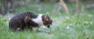
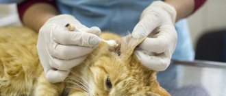
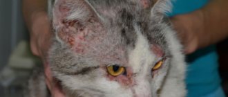
If a cat has an ulcer on the nose, this may be a sign of pathologies such as infection with pathogenic microflora, an allergic reaction, polyps, or traumatic injuries. In addition to wounds, the condition may be accompanied by fever, sneezing, coughing, and changes in the animal’s behavior. At the first symptoms, it is recommended to take your pet to a veterinarian, who will prescribe medications or perform surgery.
According to statistics, timely treatment of calicivirus saves the lives of 80% of animals.
Treatment of necrosis in cats
If necrosis is detected in an animal, a course of treatment should be started immediately, since in the early stages of the disease there is a possibility of a complete cure without surgery. In the final stages, surgery cannot be avoided. After surgery, the veterinarian will prescribe a course of strong antibiotics that are injected directly into the affected bone.
A wound is an open mechanical damage to the skin, mucous membrane, as well as underlying tissues and organs, characterized by gaping, pain, bleeding, and sometimes dysfunction.
Minor damage to the integrity of only the epidermis is called abrasions or scratches. Veterinary specialists distinguish between wound edges, walls, bottom and cavity. The edges of the wound are formed by skin, the walls - by muscles and fascia and the loose connective tissue located between them. The bottom is the deepest part of the wound. The space between the walls of the wound is called the wound canal. Puncture wounds usually have a deep wound channel, but superficial damage to the channel does not. The space between the edges is called the wound opening. The shape and size of the wound opening depend on the nature of the wound. The wound opening can be oval, round, triangular, star-shaped, crescent-shaped, etc.
In the case when, as a result of a wound, any part of the body is completely pierced, then such a wound is called a through wound. In this case, a distinction is made between the inlet and outlet openings. If a wounding object perforates the wall of an anatomical cavity (peritoneum, pleura), the wound is called a penetrating wound. Such wounds most often have only one entry hole.
Since mechanical open tissue damage causes a general, often severe pathological reaction in a cat, they speak of wound disease. Wound disease is a symptom complex of local and general neurohumoral disorders in the body caused by injury and the subsequent development of toxic-infectious processes.
Depending on the nature of the wounding object and the mechanism of action, several types of wounds are distinguished. There are many similarities between different wounds, and each form has its own characteristics.
A puncture wound occurs in a cat when long pointed objects (nail, needle, stick, etc.) are inserted into the tissue. The nature of tissue damage depends on the shape of the object. Punctured objects with a sharp end easily push tissue apart; with rough surfaces, tissues are torn, crushing and crushing them along the wound channel. A puncture wound communicating with one or another anatomical cavity is called a penetrating wound. Puncture wounds are deep. The gaping is weak or may be absent. If the large blood vessels are not damaged, then the bleeding is small, sometimes it appears a few minutes after the injury. It must be borne in mind that with a penetrating wound, external bleeding is sometimes absent, while at the same time blood accumulates in the damaged anatomical cavity (intracavitary bleeding).
An incised wound is formed when tissue is cut with sharp objects (glass, iron, etc.). An incised wound has smooth edges and walls and is accompanied by a slight gaping, especially in the middle part of the wound and bleeding. With proper and timely treatment, such wounds heal relatively quickly.
A lacerated wound in a cat is formed when tissue is torn by sharp-pointed objects acting in an oblique direction (the claws of other cats, barbed wire, tree branches, etc.). The wound channel is formed of varying depths and has sinus-shaped branches. The edges are uneven, sometimes jagged, with a significant separation of the skin hanging down in the form of a flap. The cat's pain reaction is very pronounced at the time of injury, then gradually decreases. The gaping is very pronounced; bleeding is insignificant or absent altogether, which is explained by the uneven rupture of the vessel walls. Lacerated wounds are characterized by prolonged healing and tissue necrosis. In such wounds there is a favorable environment for the development of wound infection and the spread of the purulent process.
A bite wound is caused by the teeth of other cats, dogs, and wild animals. A bite wound has a variety of shapes; the damage depends on the depth of the teeth and the nature of the jaw movement, as animals sometimes try to tear out a piece of tissue. Dogs tear skin and muscles, leaving puncture wounds on the body from fangs, sometimes such a wound is accompanied by broken bones. A bite wound almost always festeres as a result of saliva and plaque from the teeth entering the wound canal. Pet owners should keep in mind that if bitten, especially by wild animals, your cat can become infected with a dangerous disease such as rabies (rabies in cats).
A bruised wound in a cat occurs as a result of the impact of a blunt object, at the site of which we observe a rupture of the skin, severe bruising of muscles, nerves and other tissues, sometimes there is a broken bone and slight bleeding. A strong pain reaction at the time of injury soon weakens, as the nerve endings temporarily lose their ability to conduct impulses (wound stupor). Sometimes a traumatic object in a cat tears off only the epidermis, leading to rupture of the papillary layer of skin. During a clinical examination of such areas, a veterinarian will detect bruises and abrasions. Deprived of blood supply and innervation, muscles provide a good breeding ground for the development of wound infection and purulent-putrefactive inflammation in the tissues adjacent to the wound canal.
A crush wound in cats occurs under the influence of significant force and pressure from a wounding object, pinching or strong compression of tissue with crushing at the moment of collision with the object. Damage in a cat has the features of gross anatomical destruction; tissues and organs are crushed and soaked in blood; fragments of fascia and tendons hang from the wound. There may be no bleeding in such wounds, as vascular thrombosis quickly occurs. Due to the large destruction of soft tissues and hemorrhages, necrotic foci are created in which wound infection quickly develops. Hence, crush wounds require urgent surgical treatment.
Symptoms of wounds.
Each wound is characterized by the following clinical signs - pain, disruption of the integrity of the integument, bleeding, gaping, sometimes dysfunction of the damaged organ, swelling.
The cat's pain occurs instantly. At the moment of injury and over time it gradually decreases. At the same time, increased local inflammation in the wound increases pain and vice versa. A decrease in the inflammatory response leads to a decrease in pain in the animal.
The more abundant the sensitive innervation of the tissue (skin of the genital organs, cornea, peritoneum, periosteum), the stronger the pain reaction in the cat. Injury to parenchymal organs does not cause severe pain. The intensity and duration of the pain reaction depends on the location of the wound, the nature of the damage and individual reactivity. The cat is a very sensitive animal to pain and can sometimes die from pain shock.
Clinically, the pain reaction in a cat is manifested by an acceleration of heart contractions and dilation of the pupils. The cat develops a defensive reaction when palpating the wound area - trying to bite the owner or veterinarian.
It must be borne in mind that painful irritations adversely affect many functions of the body: secretory, motor function of the gastrointestinal tract, lymph and blood circulation, cardiac activity, fat, protein and vitamin-mineral metabolism, etc.
Violation of the integrity of the integument. With puncture and bite wounds, owners cannot always immediately detect the entrance hole, finding only wet fur.
The bleeding that occurs when a cat is wounded depends on the nature of the damaged blood vessel and the type of wound itself. Bleeding in a cat can be external, internal, arterial, venous, capillary, parenchymal and mixed. By time of origin: primary and secondary, by frequency – single and repeated.
External and internal bleeding. External bleeding is manifested in a cat by the outpouring of blood from a wound into the external environment. Recognizing external bleeding is not difficult. With internal bleeding, blood flows into damaged tissue or anatomical cavity (peritoneum, pleura, joint, stomach, etc.). In this regard, a distinction is made between interstitial and intracavitary bleeding. Recognizing internal bleeding is often difficult. Internal bleeding is characterized by weakening and increased pulse rate, pallor of visible mucous membranes, general weakness and shortness of breath.
Primary bleeding in a cat usually occurs immediately following an injury. Secondary, or recurrent, bleeding occurs several hours or days after the primary bleeding has stopped. The cause of secondary bleeding can be: insufficiently thorough stopping of primary bleeding, rough change of dressing, damage to blood vessels from bone fragments, foreign bodies not removed from the wound. The cause of secondary bleeding may be a violation of thrombo- and fibrin formation due to a lack of vitamins C and K in the body; complication of a wound by infection, especially putrefactive infection, in which melting of tissues and vascular blood clots occurs, destruction of the vascular wall by a malignant tumor.
Prevention of secondary and recurrent bleeding in a cat consists of timely and complete surgical treatment of the wound, removal of foreign bodies located near large vessels, and careful stoppage of primary bleeding. Drugs that increase blood clotting are used. It is necessary to eliminate conditions favorable for the development of wound infection. For this purpose, dead tissue is carefully removed by surgical, physicochemical or enzymatic means; suppress infection with antibiotics, sulfonamide drugs and other antimicrobial drugs. The wound must be provided with complete rest, while preventing its re-traumatization.
Wound healing.
Wound healing in animals can occur by primary and secondary intention.
Primary intention is the most advanced wound healing process, in which fusion of the edges and walls occurs with mild aseptic inflammation, slight swelling and the formation of a minimal scar. Healing by primary intention is possible only with a tight and correct connection of the edges and walls of the wounds, a minimum amount of dead tissue, stopping bleeding, the absence of foreign bodies and infection, i.e. subject to careful asepsis and antisepsis. Clean surgical and fresh casual wounds heal by primary intention after appropriate treatment (use of chemicals, antibiotics, surgical excision of dead tissue, removal of foreign bodies).
Healing in the wound begins in the first hours, after bleeding stops and the edges of the wound come together: hyperemia develops, the reaction of the wound environment changes in an acidic direction, a thin layer of fibrin falls on the wound walls, gluing the edges of the wound (primary adhesion). During the first day, the wound gap quickly fills with migrating leukocytes, lymphocytes, fibroblasts, polyblasts, and macrophages.
Secondary intention is the healing of a wound through the development of granulation tissue, followed by epithelization and scarring. Healing by secondary intention takes a long time in the animal. Healing by secondary intention occurs in cases where the wound does not have conditions for primary intention (wound gaping, the presence of dead tissue in the wound, the development of wound infection, purulent inflammation, repeated bleeding and contamination). With secondary intention, after a few hours the wound is covered with a small layer of fibrin and wound secretion. After 2 days, the wound secretion becomes somewhat transparent and pus is released. On the 3-4th day, granulation tissue (small reddish grains) forms in the wound, where there are no dead cells. Subsequently, granulation tissue covers the entire wound cavity. Over time, granulation tissue becomes connective and later scar tissue.
Healing under the scab. Healing under a scab in cats occurs after cutting shallow wounds to the dermis. The blood that flows out, dries, glues the edges of the damaged wound and forms a scab, under which healing occurs.
Treatment of wounds in cats.
When treating a wound in a cat, animal owners should strive to prevent infection in the wound and speed up the healing process by any available means.
Different types of wounds sustained by a cat require different approaches to wound care.
When providing first aid, you will need to cut the hair around the wound, lubricate the edges of the wound with a solution of iodine and brilliant green. In the case where there is a small cut or scratch, it will be enough to wash the wound with a 3% solution of hydrogen peroxide or a light pink solution of potassium permanganate.
If there are foreign objects in the wound (glass, sand, etc.), they must be removed with tweezers, after which the wound cavity should be rinsed several times with a 3% solution of hydrogen peroxide or a weak solution of potassium permanganate. Then lubricate the skin around the wound with brilliant green, iodine, alcohol or vodka. We powder the wound with tricillin powder, streptocide, ranosan, etc.
If the wound is deep, gapes greatly and you are not able to properly treat it, then cover the wound with a sterile napkin (sterile bandage), apply a bandage and contact a veterinary clinic as soon as possible, where all the necessary medical procedures will be carried out.
If a limb is injured and there is bleeding, it is necessary to apply a braid or bandage above the wound site to stop the bleeding. For minor bleeding, apply a tight bandage.
If the abdominal cavity is injured, the cat should not be given water; in case of bleeding from the lungs, only cold water can be given; If there is bloody vomiting, you should never feed the animal. It is urgently necessary to contact the nearest veterinary clinic.
If a cat has bite wounds, it is necessary to treat such a wound especially carefully. Because when bitten, saliva, hair and dirt from the skin get into the wound. It is recommended to thoroughly wash such wounds with a 3% solution of hydrogen peroxide from a disposable syringe.
In order to speed up wound healing, it is recommended to use antibiotics, vitamins, enzymes, etc.
Keeping and caring for an injured cat.
At home, caring for a wounded cat should consist of preventing it from scratching or biting treated wounds, stitches, or bandages. Sometimes you have to resort to using a special collar for this. The dressings applied to the wound must be dry and clean and changed according to the recommendations given by the veterinarian.
Dangerous conditions include necrosis in cats. There are several types of tissue necrosis, which are accompanied by changes in skin color, the appearance of non-healing ulcers, and an unpleasant odor. The necrotic process is irreversible, so at the first symptoms, the owner should take the cat to the veterinarian, who will prescribe treatment and give preventive recommendations.
According to veterinarian statistics, 99% of animals with gangrene require amputation of the affected limb.
Further care
After self-treatment of the wound or a visit to the veterinarian, the healing of the kitten’s wound depends solely on proper care for it. “Murkosha” advises all owners to be patient, as this process takes up to two weeks. To speed up your pet's recovery, follow these tips:
- Do not allow the cat to lick wounds. He will instinctively want to do this, but this will not heal them. To prevent this behavior, veterinarians advise using a special collar that will prevent the cat from scratching and licking the wound. — If your pet has dressings, they must be changed regularly when they become dirty and always kept dry. If you cannot change the bandages yourself, then ask friends for help or go to the clinic for bandaging. — The cat must take all prescribed medications on time. The treatment plan will be drawn up in advance by the veterinarian, and all you have to do is strictly follow it. If this is not done, the bacteria will become resistant to the drug and the treatment will be in vain. — Not only the use of medications, but also a balanced diet can speed up your pet’s recovery. The cat should be fed more often than usual - about 5 times a day. The cat's diet must include protein, as it helps healing. In addition, food should be rich in vitamins. All these requirements are met by professional super-premium and holistic-class food. — Report any deterioration in your pet’s condition to your doctor immediately.
Why does necrosis occur?
The pathology is characterized by irreversible tissue death and cessation of cell activity due to impaired blood circulation, nerve conduction, and the activity of anaerobic or pyogenic bacteria. Most often, areas of the body where the blood vessels are small are affected - ears, paws, muzzle, tail, tongue. Often internal organs are affected - intestines, stomach, kidneys, liver. Depending on the location, the causes of necrosis are as follows:
- Internal organs: entry of foreign objects into the gastrointestinal tract;
- helminthiases;
- cirrhosis;
- volvulus.
- frostbite;
- glossitis (inflammation of the tongue);
Allergies or exposure to toxic chemicals can lead to tissue death.
In addition, the following factors cause tissue death in any area:
- autoimmune failure;
- allergy;
- prolonged inhalation of pesticides by animals;
- heart attack;
- electrical injury;
- disturbance of nerve conduction.
Return to contents
Calcivirosis
Calcivirosis is a dangerous viral disease of cats. In addition to the nose and oral cavity, the infection can affect the respiratory, hematopoietic and nervous systems of animals, and the digestive organs. The disease is often severe, with symptoms appearing within 1-3 weeks, and about a third of those affected die. Survivors are able to shed the virus into the environment for up to 12 months. With stress, deterioration of living conditions, infection with VLK or FIV, a relapse of calicivirus occurs. If a secondary infection occurs, the mortality rate increases to 80%.
To avoid death, you need to show the animal to a veterinarian as soon as possible.
Symptoms: how to recognize the disease?
Necrosis of internal organs in a cat develops mainly against the background of other diseases, so signs of the underlying disease are observed. The presence of tissue death is indicated by the following symptoms:
- stool disorder;
- increased blood pressure;
- heart rhythm disturbance;
- refusal to eat;
- periodic increase in temperature;
- difficulty breathing or shortness of breath;
- lethargy, weakness.
On the external parts of the body the symptoms are pronounced:
- local increase or decrease in temperature;
- bluishness or redness, and eventually blackening of the skin;
- sweetish-putrid smell;
- decreased or absent sensitivity;
- hair loss in the affected areas;;
- persistent swelling, ulcers;
- hyperthermia;
- weakness, deterioration of health;
- pain, swelling and limited mobility if the bone is affected.
Return to contents
Diagnostics
In general, diagnosing a weeping wound does not cause any difficulties; everything is quite obvious, based on the visual signs of the pathological process. It is much more important to find out which microbe caused the inflammatory process. To answer this question, it is necessary to take a sample of pathological material from the wound and inoculate it on a nutrient medium.
In addition, it may be necessary to take blood and urine tests when there is a suspicion of the endogenous origin of a weeping wound (that is, to identify hormonal and other disorders).
Treatment: what to do with necrosis?
Tissue necrosis in cats can be cured by surgery. To prevent the spread of cell death due to bacterial infection, antibiotics are effective, which the veterinarian prescribes individually. To dry out necrosis, disinfect and identify clear boundaries of the lesion, dead tissue is smeared with brilliant green or Chlorhexidine. To prevent the cat from licking the medicine and getting poisoned, the limb should be bandaged and a special collar should be put on the neck.
Necrotic areas on internal organs are excised or a complete ectomy is performed. External lesions are treated by necrectomy, in which areas of necrosis in the cat are removed with a small inclusion of healthy tissue. In case of wet type of pathology or gangrene, amputation of the limb is performed. After the stump has healed and scarred, a prosthesis is installed.
Wounds and allergies
The cause of sores on the nose may be an allergic reaction. Incorrectly selected food, various medications, vitamins, and indoor plants can lead to allergies, accompanied by rashes and itching. The cat licks the itchy area of the nasal planum with its hard, rough tongue, tearing off the thin and delicate top layer of skin. It is necessary to find the allergen and remove it from the apartment. If this is a drug, you should consult your veterinarian about replacing it. A specialist may prescribe antihistamines.
Prevention
To avoid external necrosis in a cat, veterinarians recommend not leaving your pet in the cold, protecting it from burns, and putting away household chemicals or dangerous objects that the pet can swallow (rubber bands, ribbons, New Year’s rain, polyethylene). Exposed wires should not be left unattended if the house is undergoing renovations. Even minor wounds and scratches should be treated promptly, and deeper cuts or bites should be treated under the supervision of a veterinarian. The owner should carefully care for the pet after abdominal surgery and monitor the condition of the sutures. All systemic diseases in cats must be treated promptly. It is also recommended that the cat be vaccinated and dewormed in a timely manner.
Nose injuries
The nasal planum is an organ that provides the domestic predator with a significant amount of information about the surrounding world. When getting acquainted with something new, a cat carefully sniffs the object. She does the same with new pets or guests. By touching the owner’s hands and shoes and his things with his nose, the animal “reads information” about where the person has been, with whom he has been in contact, and what he has touched.
By sniffing food, the little hunter determines not only its freshness and edibility, but also its temperature. Injuries to the nasal planum are possible both in a pet that is allowed outside and in a couch potato.
A sore on a cat’s nose can appear after a fight with relatives, an unsuccessful jump from a height, or sniffing thorny indoor plants. Pets that live in the same house from a young age often do not perceive each other as links in the food chain. They play, communicate with each other, and not always neatly.
In case of a minor injury or ulcer on the nose, the cat can be treated by lubricating the nasal planum with vegetable oil, such as sea buckthorn or olive. A little tea tree essential oil is first added to olive oil (2-3 drops per 1 tsp), which improves wound healing and helps suppress the activity of microorganisms (bacteria, viruses, fungi). Before the procedure, you can lightly wash the wound by touching it with a cotton pad soaked in a pale pink solution of potassium permanganate (potassium permanganate).
If there is no effect from home treatment within 3-5 days, a veterinarian will help cure the small predator. Perhaps an infection has joined the injury or there is an allergic reaction to something.

