The heart of four-legged pets suffers from various diseases just like humans. Among the well-known diseases of the cardiovascular system of animals is HCM (hypertrophic cardiomyopathy). In cats, this pathology is dangerous due to severe, sometimes incompatible with life, complications. The article is devoted to a detailed overview of the disease and methods of its treatment.
HCM in cats: symptoms, treatment
Feline heart disease
Pathologies occur both congenital and acquired. All existing diseases are classified according to location and cause of occurrence:
- Heart defects.
- Tissue mutations.
- Disorders of heart rhythm and conduction of electrical impulses.
- Parasitic origin.
- Inflammatory. They develop in organ tissues. The following diseases are distinguished by location:
- pericarditis;
- myocarditis;
- endocarditis.
Among these problems, cardiomyopathy develops most often in cats.
Structure of a cat's heart
Hypertrophic cardiomyopathy is the most common heart disease in cats. Interview with a cardiologist
We talked about the most common cardiac disease in cats with the head of the cardiology department, veterinary cardiologist, candidate of biological sciences Vladislava Konstantinovna Illarionova.
— Do cats have heart disease?
- Yes, there are. A cat's small heart can indeed be unhealthy, and the most common pathology is a chronic heart muscle disease called cardiomyopathy. In fact, cardiomyopathies are a whole group of myocardial diseases that slowly progress and in some animals lead to chronic heart failure, blood clots and sudden death. Heart disease is one of the ten most common causes of death in cats.
— What heart disease is most common in cats?
— Hypertrophic cardiomyopathy is the most common form of feline cardiomyopathy. The disease is characterized by thickening of the heart muscle, which is called “hypertrophy”. This is a progressive pathology that hardens the walls of the heart, reducing the ability of the ventricular chambers to effectively draw in and eject blood. In addition, such changes in the myocardium can affect the functioning of the heart valves, causing incompetence that aggravates the pathological process. Often, transformations of the walls of the heart lead to the formation of fast flows in the chambers and vessels, which cause the characteristic noise heard with a stethoscope. With the development of heart failure, the body compensates for the circulatory deficiency by increasing the frequency of heart contractions, as well as retaining salt and water. For some time, such adaptation mechanisms maintain normal blood flow, but over time, the body's resources are depleted and clinical signs of the disease begin to develop.
— How common is this disease?
— Hypertrophic cardiomyopathy is detected in approximately 15% of cats in the general population. In older cats, the prevalence reaches 29%. Most cats with hypertrophic cardiomyopathy are asymptomatic, with an incidence of cardiac death of approximately 23% within 5 years, regardless of age at diagnosis.
— What is the reason for the development of the disease?
— Most often we call such diseases idiopathic, that is, without a clear cause. However, more and more data support the genetic nature of the pathology. Here, however, it is important to say that the possible reasons may be heterogeneous. Similar changes develop in the heart secondary to high blood pressure (which is not uncommon in cats) or chronic pathology of the thyroid gland. In addition, in cats, due to stress, a pathological process in the heart can acutely occur, which is called transient myocardial hypertrophy and is most likely associated with an inflammatory reaction of the heart muscle. Such changes are sometimes recorded in cats after general anesthesia, changes in their usual lifestyle, moving, the appearance of new members in the family, and so on. Clinically, this is manifested by shortness of breath and apathy, which usually occurs 3-5 days after the event.
— Which cats are predisposed to this disease?
— Although most cats with hypertrophic cardiomyopathy are mongrel, some breeds have an increased risk of developing the pathology. These include Maine Coons, Ragdolls, British Shorthairs, Persians, Bengals, Sphynxes, Norwegian Forest cats and Burmese. However, data on the prevalence of the disease in breeding animals of different breeds is not complete. Two contractile protein gene mutations have been identified in the myosin binding protein C (MyBPC3) gene. The estimated prevalence of the MyBPC3-A31P mutation in Maine Coon cats is 35% to 42%, which is significantly higher than the prevalence of the disease in this breed. Maine Coons homozygous for this mutation, ragdolls homozygous for the MyBPC3-R820W mutation, and kittens of affected animals are at risk of developing hypertrophic cardiomyopathy.
— If the hereditary nature of the disease is proven, is there a test for early detection of pathology?
— Genetic testing for the MyBPC3-A31P and MyBPC3-R820W mutations is recommended for Maine Coon and Ragdoll cats. Tests are carried out on animals involved in breeding in order to reduce the frequency of mutations in these breeds. It is recommended to remove from breeding cats homozygous for these mutations, but leave the possibility of crossing heterozygous animals with cats negative for mutations if they have outstanding breed characteristics. Genetic tests can also be used in non-breeding Maine Coon or Ragdoll cats to determine the relative risk of developing the disease. However, Maine Coon and Ragdoll cats that test negative for the MYBPC3 mutation also have hypertrophic cardiomyopathy and, therefore, the only reliable diagnostic method is regular echocardiographic examination. For cats of other breeds, genetic testing of the MYBPC3-A31P and MYBPC3-R820W mutations is not recommended, since these mutations have been identified only in Maine Coon and Ragdoll animals.
— What symptoms are typical for heart disease in cats?
— Chronic heart failure is the most common cause of clinical signs in cats with hypertrophic cardiomyopathy. In second place is arterial thromboembolism. Sudden death without clinical manifestations is recorded in a small number of patients. There are two main mechanisms for the development of symptoms. The first is associated with an increase in pressure in the veins of the lungs due to the fact that blood is poorly pumped by the heart - this leads to the sweating of its liquid part into the lung tissue. This condition is called “pulmonary edema” and can lead to the death of the animal. The main symptom is shortness of breath. The frequency of inhalations exceeds 30 per minute at rest or during sleep, movement of the chest becomes noticeable when breathing, and sometimes the mouth opens slightly. Often such signs are accompanied by depression, reluctance to play, communicate, and eat. The cat finds a secluded place and does not agree to be persuaded to leave. Pulmonary edema is often accompanied by the accumulation of free fluid in the chest, which further aggravates the condition.
The second mechanism for the development of symptoms is associated with the formation of blood clots in the cavity of the enlarged left atrium. Such blood clots form as a result of disruption of blood flow through the heart and can reach significant sizes. The greatest danger arises from the leaching of these clots into the systemic circulation. If a thrombus goes into the vessels of the systemic circulation, then the symptoms depend entirely on its diameter and location. Most often, the thrombus stops at the site of bifurcation of the abdominal aorta into the vessels of the pelvic limbs. This causes a sudden loss of support on the hind legs, severe pain and fear. The cat cannot get up and screams pitifully. This condition is very life-threatening and the prognosis is largely determined by the diameter of the blood clot and the time between the onset of symptoms and the start of treatment. If more than 4 hours pass from the moment of the first manifestations, the prognosis worsens significantly.
— How can this disease be diagnosed?
— The main method for diagnosing feline cardiomyopathies is echocardiographic examination (ultrasound of the heart). This is a method that provides comprehensive information about the structure and function of an organ. There are a number of clear criteria for identifying pathology, which are based on the anatomical landmarks of the thickness of the walls of the heart, the shape of the thickenings, the nature of the movement of the heart muscle, and the parameters of myocardial relaxation. In addition, there are signs by which the doctor can judge the risk of pulmonary edema and the presence of free fluid in the chest. Echocardiography is a very comfortable and safe method for our patients. It does not require anesthesia and is not associated with unpleasant sensations, so most cats tolerate it well. The study lasts about 20 - 30 minutes (sometimes a little more or less), after which the doctor carries out the necessary measurements and calculations and fills out a study protocol.
It must be said that cardiologists often do not limit themselves to one research method, but carry out diagnostics in a comprehensive manner. Therefore, after echocardiography, we proceed to an electrocardiographic study (ECG), which provides invaluable information about the heart rhythm. The condition of the lungs, respiratory tract, and vessels of the chest can be assessed using an X-ray examination, and if a detailed layer-by-layer view of all structures of the chest organs is necessary, computed tomography is performed. Another important step in diagnosing heart disease is measuring blood pressure. If the doctor still has doubts about the cause of the symptoms and he needs to differentiate the respiratory and cardiovascular systems according to the degree of importance of their influence on the formation of the clinical picture, then there is a diagnostic method such as determining the concentration in the blood or pleural fluid of natriuretic peptide, which is called a cardiac marker and increases with significant heart failure. Another auxiliary test is to determine the concentration in the blood of another cardiac marker - cardiac troponin I, which increases with the destruction of the myocardium and provides information about acute pathologies of the heart muscle, for example, such as transient myocardial hypertrophy (myocarditis).
— What is rapid echo screening?
“There are situations when life depends on emergency treatment measures. In this case, the doctor can simply "take a look" at the heart by performing a quick ultrasound examination of the chest. To do this, the cat is usually not fixed on its side, but screening is carried out in positions that are more comfortable for the animal - on the chest, standing or sitting. The fact is that if there is pulmonary edema or fluid accumulation in the chest, keeping the animal in a position on its side can cost it its life. Therefore, the doctor’s task is to quickly and with the least stress for the cat conduct a study for the presence of pulmonary edema, pleural effusion, and significant changes in the heart. This is necessary for choosing treatment tactics in the acute period and does not replace a full echocardiographic study after stabilization of the animal’s condition.
— Is it possible to cure the disease?
“Unfortunately, the vast majority of cardiomyopathies only change in the direction of worsening. The only exception is transient myocardial hypertrophy, which may completely resolve after a few months. However, treatment can prolong life and improve quality of life in patients with clinical signs. The main drugs for chronic therapy are diuretics, inhibitors of hormonal systems involved in the formation and development of heart failure, drugs that reduce the risk of thrombosis, antihypertensive and antiarrhythmic drugs, as well as drugs that improve the functioning of the heart muscle and dilate the blood vessels of the lungs.
— What do you need to know to properly care for a sick animal at home?
— A very important factor for cats is a calm and comfortable environment. Any changes in the usual environment have a bad effect on a sick animal. So you shouldn’t take other animals, change the cat’s diet or habitual lifestyle. It is very important to follow the medication administration schedule. Over time, this becomes a habit for both the owners and the patient. A great help for monitoring the condition of the animal is to count the frequency of respiratory movements. This procedure is usually carried out when the cat is calm or sleeping. To count, take a minute and record the number of breaths. The upper limit of normal is 30 breaths per minute, although in cases of severe chronic illness the frequency may be slightly higher. In any case, this should definitely be discussed with your doctor. In addition, it is necessary to discuss with the doctor the schedule of repeated examinations and the possibility of communication in emergency situations.
Cardiomyopathy: etiology and pathogenesis
Feline hypertrophic cardiomyopathy (HCM) is a pathology accompanied by hardening of the walls of the ventricle. As the disease develops, the volume of the heart chambers is significantly reduced, subsequently provoking heart failure. Timely treatment helps prevent death, but in advanced forms of the disease the prognosis is poor. The disease mainly manifests itself early in the pet's life - at the age of 6 months.
Animals with genetic heart abnormalities do not survive to puberty
HCM is considered a relatively new disease. Research into this pathology was actively carried out at the beginning of the last decade in the USA.
Scientists have concluded that breeds most susceptible to the disease include Ragdoll, Sphynx , Norwegian Forest, Scottish Fold , Persian and Abyssinian breeds. In Maine Coons, transmission of the disease at the genetic level has been confirmed. Most often the disease is observed in males.
The most susceptible breeds to the disease
Among such mass-bred breeds as Siamese , British Shorthair , Siberian and Russian Blue, no hereditary predisposition has been identified. Representatives of these varieties suffer from secondary forms of pathology.
What happens to the heart with HCM?
The clinical picture of a typical cardiomyopathy is characterized by the following manifestations:
- Reduction in the size of the left ventricle, which can lead to a decrease in the amount of blood supplied. In addition, the mobility of the walls decreases. This leads to the release of blood into the vessels of the lungs.
- As the disease progresses, the left atrium enlarges. The resulting blood clots contribute to impaired hemostasis, leading to paralysis of the hind limbs.
- Further, the mitral valve is involved in the pathological process. As a result of a change in its location, an obstruction occurs in the exit of blood into the aorta. This hitch in the blood flow is accompanied by a systolic murmur.
- Myocardial compaction causes blood stagnation in the atria and malfunction of the atrioventricular valve. As a result, aortic obstruction and circulatory deficiency are noted.
Image of a heart affected by pathology
Types of HCM
Hypertrophic cardiomyopathy of furry pets is divided into 2 types: primary and secondary. The origin of the first type, which occurs in obstructive and non-obstructive forms, is currently under study. The second (acquired) develops under the influence of another disease.
Table 1. Types of HCM in cats
| Type | Characteristic |
| Obstructive | Due to the expansion of the myocardium, the pressure in the left ventricle increases, which increases the rate of blood flow into the aorta. As a result, the bicuspid valve leaf opens and closes involuntarily |
| Non-obstructive | The mechanism of manifestation of the pathology is identical to the previous one, except that high blood flow speed does not affect the functioning of the valve |
| Acquired | It is provoked by existing diseases and age-related transformations, accompanied by mutations in the structure of the myocardium. Infectious pathologies, disruptions in the endocrine system, injuries and toxins are the triggering factors. |
The primary type of the disease, which is characterized by genetic origin, manifests itself before 5 years. The secondary form occurs mainly in older cats after 7 years.
The secondary form of the disease occurs in adult cats
Possible complications
The progression of hypertrophic cardiomyopathy affects all parts of the heart and the circulatory system as a whole. This occurs due to the overcrowding of the lung vessels with blood. Due to slow blood circulation in the cat's heart chambers, blood clots form.
The danger of HCM is the formation of blood clots
The mechanism of pathology development
Hypertrophy of the walls of the ventricles significantly impairs the functioning of the heart, which leads to stagnation of blood in the atria. Weak blood flow causes circulatory hypoxia (oxygen starvation caused by poor circulation). Due to spasm of peripheral vessels, the risk of thrombosis increases. If the disease is not treated, it can lead to the development of heart failure, dangerous arrhythmias, and even cause the death of the animal.
Depending on the location, HCM is classified into the right or left ventricle. In cats, lesions of the left ventricle and interventricular septum are more common.
The pathological process slows down the growth of muscle tissue that forms the contractile system of the heart. The body compensates for this deficiency with connective tissue, which thickens the walls of the myocardium. Peculiar scars appear on the heart, reducing the size of the heart chambers. For the same reason, the elasticity of the organ decreases.
Symptoms of the disease
The main manifestations of hypertrophic cardiomyopathy are poor exercise tolerance and shortness of breath. If the disease is asymptomatic, there is a high probability of death due to thromboembolism, which develops as a result of pulmonary edema.
Dyspnea and paralysis are common in HCM
The main signs of HCM are the following symptoms:
- Physical state. An exhausted animal loses interest in active games and sleeps a lot.
- Breath. Severe, accompanied by wheezing and gurgling. Under heavy load, the pet experiences difficulty in inhaling. This is caused by slowing down the movement of blood in the veins. The cat breathes with its belly, sticking out its tongue.
- Dyspnea. Attacks of suffocation often lead to fainting as a result of lack of oxygen.
- Character of the pulse. Filiform.
- Condition of mucous membranes. Due to oxygen starvation, the shells acquire a blue tint.
- Cough. Reflex, caused by excessive pressure of an enlarged heart on the trachea. To cope with an attack, the cat sticks out its neck, spreading its forelimbs for greater ventilation.
- Hydrothorax and ascites. The release of exudate contributes to the formation of swelling in the peritoneum and chest.
- Paralysis of the hind limbs. If a blood clot blocks the lumen in the vessels, the animal’s legs in the pelvic area fail.
- Body mass . Poor weight gain.
- Heart murmurs and tachycardia.
If you notice any of the symptoms, take your pet to the veterinarian
Symptoms of cardiomyopathy
Unfortunately, cardiomyopathy can occur for a long time without any symptoms, and then manifest itself with symptoms common to many cardiac diseases:
- shortness of breath with minor physical effort, walking;
- arrhythmias;
- pain in the heart area;
- constant loss of strength;
- dizziness;
- fainting states.
Often, cardiomyopathy can be suspected only when the disease is already in the stage of decompensation and all the symptoms of heart failure are present:
- shortness of breath even at rest;
- presence of swelling in the legs;
- ascites (accumulation of fluid in the abdominal cavity), enlarged liver, etc.;
- atrial fibrillation and other types of arrhythmias;
- cough that gets worse when lying down;
- attacks of suffocation;
- pulmonary edema.
1 Ultrasound of the heart for cardiopathy
2 ECGs for cardiomyopathies
3 Blood tests for cardiomyopathy
Reasons for the development of the disease
The main cause of the disease is considered to be a gene mutation. The pathology is inherited among some cat breeds. American experts concluded that the gene responsible for myosin binding protein is to blame. Maine Coons and Ragdolls are predominantly affected by this gene mutation. It has been proven that more than 10 genes are involved in the development of the disease.
Representatives of the Maine Coon breed are more susceptible to pathology than others
In addition to breed predisposition, a number of other reasons influence the development of cardiomyopathy.
Table 2. Provoking factors
| Provoking factor | Characteristic |
| Congenital myocardial pathologies | Any diseases occurring in this part of the heart are fraught with hardening of its walls |
| Infusion therapy | If the rate of intravenous penetration of the drug into the pet’s body does not correspond to its physiology, the pressure in the vessels increases. As a result, stagnation occurs, leading to swelling of the lungs. Insufficient oxygen supply leads to hypoxia |
| Malignant neoplasms | The formation of lymphoma is fraught with mutation of the myocardial structure |
| Endocrine diseases | Increased activity of the thyroid gland contributes to the development of tachycardia. Due to increased production of growth hormone, the walls of the heart muscle thicken |
| Taurine deficiency | This amino acid, essential for cats, regulates organ contractility, has an anti-ischemic effect and maintains the integrity of cell membranes. Deficiency of this element destabilizes the heart muscle |
| Application of anesthesia | Pets may develop hypertrophic cardiomyopathy after being under general anesthesia. The disease manifests itself either immediately or several days after artificial immersion in sleep |
| Chronic intoxication | Overdose of medications, chemical poisoning, products of pathogenic activity of parasites negatively affect the fibers of the heart muscle and contribute to the development of ventricular hypertrophy |
| High blood pressure | Constant deviation from the norm provokes wear and tear of the organ |
Causes of pathology
The primary form of the disease is provoked by genetic mutations that occur during fetal development. Secondary pathology occurs under the influence of a number of predisposing factors, such as:
- poor nutrition;
- endocrine disorders;
- lack of taurine;
- high blood pressure;
- oncological diseases;
- severe intoxication;
- lung damage;
- congenital anomalies of myocardial development;
- general anesthesia;
- chronic infectious foci.
High blood pressure in cats contributes to the development of hypertrophic cardiomyopathy
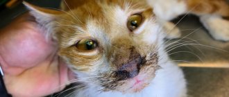
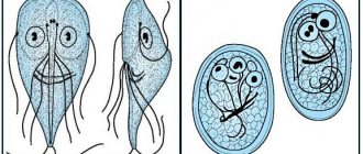
A predisposing factor in the occurrence of this heart pathology can be the constant presence of helminths in the cat’s body, especially large ones. The fact is that parasites in the process of life produce toxins that can provoke a number of pathologies. If deworming is not carried out on time, the cat will suffer from chronic intoxication, which negatively affects the functioning of the heart.
Diagnosis of the disease
The difficulty of detection is caused by the hidden nature of the pathology. Malfunctions in the functioning of the organ can be detected by listening to heart murmurs. In the presence of HCM, they resemble a gallop.
An echocardiogram is considered the most informative way to diagnose HCM
Basic methods:
- Anamnesis collection. The owner must describe the pet's condition in detail. Any deviation in behavior may signal the development of the disease.
- Echocardiography is the most accurate diagnostic method. It makes it possible to thoroughly study the structure of the heart muscle. Each cat that is at risk by breed should be examined at least once a year. Before surgery that involves the use of anesthesia, it is also necessary to check for the presence of HCM.
- Electrocardiography. This survey is not informative enough. It can be used to detect tachycardia, ventricular arrhythmia and widening of the QRS interval. However, it is not used as an independent method for diagnosing pathology.
- X-ray diagnostics. The shape and size of the heart muscle, as well as the presence of fluid in the pleural cavity, are assessed.
- Auscultation. Listening allows you to identify extraneous noise and evaluate your heart rhythm.
- Pressure measurement. With HCM, blood pressure will be elevated.
- Visual assessment. Blue discoloration of the mucous membranes indicates a malfunction of the heart muscle.
Pressure measurement is an auxiliary diagnostic method
If the animal has a severe form of hypertrophic cardiomyopathy, hardware examinations are not performed. Any manipulation negatively affects the emotional state of the pet and increases the manifestation of unpleasant symptoms.
Is there anything that can be done?
Many cat owners, upon hearing such a diagnosis, perceive it as a death sentence. Unfortunately, in some cases they actually have every reason to do so. However, this is not always the case. The main thing is to know what to do if your cat has cardiomyopathy.
© shutterstock
As practice shows, if a cat receives appropriate treatment, it may well live for quite a long time.
First of all, a good doctor will prescribe medication for a sick animal.:
- Diuretics. They are an auxiliary drug that allows you to avoid congestion, and therefore pleural effusions and pulmonary edema.
- ACE inhibitors will be needed to reduce high blood pressure and relieve heart failure.
- A calcium channel blocker will reduce your heart rate by helping to relax the overdeveloped myocardial wall. In some cases, the heart is even partially restored.
- Beta blockers help prevent arrhythmia and related complications.
Treatment of the disease
The developed treatment regimens for pathology do not make it possible to completely overcome the disease. The main emphasis is on symptomatic therapy. In severe cases, in addition to medication, special equipment is used.
Hospital treatment
The duration of stay in the clinic is 3 days. Any hardware intervention is carried out as carefully as possible in relation to the animal. Basic procedures:
- To stabilize the condition, the animal is placed in an oxygen box, which is a chamber with a constant supply of oxygen.
- After you feel better, if necessary, the fluid accumulated in the pleura is removed (thoracentesis). This helps ease the breathing process.
While in the clinic, owners are allowed to visit their pets. Such visits speed up the healing process.
One of the methods of express therapy is thoracentesis.
Drug treatment
Therapy is based on stabilizing the heart rate, preventing the formation of blood clots and relieving pulmonary edema.
Drugs used:
- Beta blockers. "Verapamil" and "Carvedilol". Reduces the load on the heart.
- Thrombolytics. "Alteplase". Blood clots dissolve.
- Antiplatelet agents. Heparin and Aspirin. Prevents the formation of blood clots.
- Diuretics. "Temisal", "Eufillin" and "Furosemide". Reduce congestion and eliminate swelling.
- Taurine-containing preparations. Thin the blood and improve the condition of blood vessels. Vitamins for cats from, GimP.
Beta blocker "Verapamil" has a positive effect on cardiac tone
For older pets suffering from secondary hypertrophic cardiomyopathy, the root cause is eliminated. Animals must be registered with a veterinarian and undergo regular examinations.
The dosage is prescribed by the doctor, based on the degree of organ damage.
Diet food
In addition to taking medications, the cat is put on a special diet that helps stabilize blood pressure. It is based on avoiding salt. This substance promotes fluid retention.
It is important to enrich the nutrition plan with the following elements:
- taurine;
- fatty acid;
- L-carnitine.
If there is a lack of vitamins, food supplements are given, sold in veterinary pharmacies.
Therapeutic diet involves avoiding salt intake
If before the onset of HCM the diet consisted of dry food, it is necessary to switch the animal to food designed for pets with pathologies of the cardiovascular system.
During treatment, the animal should not be overfed, since obesity contributes to the formation of additional stress on the heart.
Excess weight negatively affects the functioning of the heart muscle
Treatment of RCM in cats
Treatment of restrictive cardiomyopathy takes place in the hospital of the RosVet VC; the veterinary cardiologist of the clinic strongly advises hospitalizing the pet for emergency care. This is especially true for cats with acute and severe congestive heart failure. For mild or undiagnosed symptoms, treatment at home is acceptable, but in compliance with all doctor’s instructions.
Possible manipulations:
- in case of severe shortness of breath, the cat is connected to an oxygen chamber;
- thoracentesis is necessary for pleural effusion, it poses a serious threat to the life of the animal;
- if there are signs of dehydration (dehydration), sodium solutions are prescribed with caution, slowly, so as not to provoke CHF.
Stress in a cat with RCM is strictly contraindicated, so it is placed in a separate cage in the hospital and is minimally disturbed. If the body temperature is reduced, apply a heating pad. A diet with a minimum of salt is prescribed; if necessary, the cat is fed by hand.
Prescription of drugs:
- furosemide is given initially in the maximum dosage with a gradual reduction to the minimum doses;
- diltiazem lowers the pulse and works well for supraventricular arrhythmia;
- beta blockers are needed to slow the heart rate and treat arrhythmias;
- enalapril reduces fluid retention and reduces the need for diuretics;
- Digoxin is used for decreased systolic function and atrial fibrillation.
Aspirin is prescribed to prevent thromboembolism, but it is less effective than clopidogrel. It is mandatory to stabilize current pathological conditions - in case of hypothermia or dehydration (low temperature and dehydration).
Things to remember:
- diltiazem and beta blockers are not used together; their use can lead to bradycardia, hypotension and acute atrioventricular block;
- enalapril in cats with severe dehydration and low sodium levels in the blood can cause azotemia, hypokalemia and low blood pressure;
- Long-term use of aspirin increases the risk of side effects when taking enalapril, especially the kidneys.
A veterinary cardiologist at the RosVet VC advises keeping a cat with RCM in the hospital for at least 7-10 days under round-the-clock supervision. A complete re-examination is carried out on the 14th day, and if you feel stable, repeat every 2-4 months.
The prognosis for the disease ranges from cautious to unfavorable. It all depends on the stage of restrictive cardiomyopathy, the age of the cat and its general condition. Under favorable conditions and timely treatment, the lifespan of cats with RCM can be extended to 2 years, but on average - 3-12 months.
If your cat is genetically predisposed to cardiomyopathy, signs of general malaise appear, the animal is apathetic, refuses food and is breathing heavily - do not hesitate! Call by phone, 24 hours a day. Make an appointment with a cardiologist and have your pet examined to prevent the development of critical situations.
Disease prognosis
The disease development scenario is determined by the following factors:
- timely detection of the disease;
- degree of severity of symptoms;
- the presence of swelling and thromboembolism.
If the pathology is detected at an early stage, subject to proper nutrition and well-chosen therapy, the prognosis is favorable. Life expectancy is not reduced. If left untreated, the pet dies. Death occurs due to thrombosis or heart failure.
Stress can make your pet's condition worse
It is important to remember that any stress can worsen a pet’s physical condition. Therefore, it is necessary to protect the animal from emotional stress.
Prevention of HCM
The leading preventive direction is early diagnosis. Your pet should be brought in regularly for examination using echocardiography. You should also enrich the animal’s diet with proteins and taurine.
Cats suffering from cardiomyopathy do not participate in breeding.
Timely diagnosis of the disease is the best preventive measure
Hypertrophic cardiomyopathy is a common pathology among cats. Maine Coons and Ragdolls are more susceptible to the disease. Therefore, owners of representatives of these breeds should be extremely attentive to the physical condition of the pet.
What can a veterinarian do?
As mentioned above, even the best doctors usually cannot provide truly effective treatment for cardiomyopathy. The only thing they can do is subject the cat to procedures that will allow them to collect more information about the disease and give the owner recommendations that will make the animal’s life easier, giving them a few extra months or even years of life.
A chest x-ray is required. This usually makes it possible to identify the pathology of the development of the heart chambers.
Electrocardiography is also often used. It makes it possible to determine tachycardia and arrhythmia - very often it is at this stage that the diagnosis of CGMP is made.
Ultrasound of the heart is not always used, but it is thanks to this method that the most information about the form of the deviation can be collected. For example, you can determine how thick the myocardial wall is, whether there are blood clots in the arteries, and also determine the amount of blood flow.
Those cats that are predisposed to cardiomyopathy should be examined with the utmost care. This includes not only individual individuals, but also entire breeds, among which heart disease is common.